
Alar ligament
Updates to Article Attributes
Body
was changed:
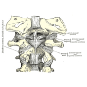
The alar ligaments join the lateral margins of the sloping upper posterior margin of the dens of C2 to the lateral margins of the foramen magnum (adjacent to the occipital condyles) and lie on either side of the apical ligament. They may be oblique or vertical and are thickest at the occipital attachment. They are paired ligaments that are very strong and limit axial rotation and contralateral lateral flexion of the head.
In conjunction with the transverse band of the cruciform ligament, they are the primary stabilisers of the atlantoaxial joint 2.
Related pathology
-<p>The <strong>alar ligaments </strong>join the lateral margins of the sloping upper posterior margin of the dens of <a href="/articles/axis-c2">C2</a> to the lateral margins of the <a href="/articles/foramen-magnum">foramen magnum</a> (adjacent to the <a href="/articles/occipital-bone">occipital condyles</a>) and lie on either side of the <a href="/articles/apical-ligament">apical ligament</a>. They may be oblique or vertical and are thickest at the occipital attachment. They are paired ligaments that are very strong and limit axial rotation and contralateral lateral flexion of the head.</p><p>In conjunction with the transverse band of the <a href="/articles/cruciate-ligament-of-the-atlas">cruciform ligament</a>, they are the primary stabilisers of the <a href="/articles/atlanto-axial-articulation">atlantoaxial joint</a> <sup>2</sup>.</p><h4>Related pathology</h4><ul><li><a title="Calcification of the alar ligament" href="/articles/calcification-of-the-alar-ligament">calcification of the alar ligaments</a></li></ul>- +<p>The <strong>alar ligaments </strong>join the lateral margins of the sloping upper posterior margin of the dens of <a href="/articles/axis-c2">C2</a> to the lateral margins of the <a href="/articles/foramen-magnum">foramen magnum</a> (adjacent to the <a href="/articles/occipital-bone">occipital condyles</a>) and lie on either side of the <a href="/articles/apical-ligament">apical ligament</a>. They may be oblique or vertical and are thickest at the occipital attachment. They are paired ligaments that are very strong and limit axial rotation and contralateral lateral flexion of the head.</p><p>In conjunction with the transverse band of the <a href="/articles/cruciate-ligament-of-the-atlas">cruciform ligament</a>, they are the primary stabilisers of the <a href="/articles/atlanto-axial-articulation">atlantoaxial joint</a> <sup>2</sup>.</p><h4>Related pathology</h4><ul><li><a href="/articles/alar-ligament-calcification">calcification of the alar ligaments</a></li></ul>
Images Changes:
Image 2 Diagram ( create )


 Unable to process the form. Check for errors and try again.