
Ameloblastoma
Updates to Article Attributes
Ameloblastomas (previously known as an adamantinoma of the jaw) are benign, locally aggressive tumours that arise from the mandible, or less commonly from the maxilla.
Epidemiology
Ameloblastomas are the second most common odontogenic tumour (odontoma is the most common) and account for up to a 3rd of
They are slow growing and tend to present in the 3rd to 5th decades of life, with no gender predilection 4.
Clinical presentation
Ameloblastomas typically occur as hard painless lesions near the angle of the mandible in the region of the 3rd molar tooth (48 and 38) although they can occur anywereanywhere along the alveolus of the mandible (80%) and maxilla (20%). When the maxilla is involved, the tumour is located in the premolar region, and can extend up in the maxillary sinus.
Although benign, it is a locally aggressive neoplasm with a high rate of recurrence. Approximately 20% of cases are associated with dentigerous cysts and unerruptedunerupted teeth.
Pathology
Ameloblastomas (not surprisingly)Unsurprisingly, ameloblastomas arise from ameloblasts, (partwhich are part of the odontogenic epithelium, responsible for enamel production and eventual crown formation).
Three variants are described:
- simple (no nodule) - best prognosis
- luminal - single nodule projecting into the cyst
- mural - multiple nodules (often only microscopic) in the wall of the cyst
Radiographic features
Plain film and CT
It is classically seen as a multilocualted (80%), expansile "soap-bubble" lesion, with well demarcated borders and no matrix calcification. Occasionally erosion of the adjacent tooth roots can be seen which is highly specific. When larger it may also erode through cortex into adjacent soft tissues.
MRI
In general ameloblastomas demonstrate a mixed solid and cystic pattern, with a thick irregular wall, often with papillary solid structures projecting into the lesion. These components tend to vividly enhance.
Treatment and prognosis
Ameoloblastomas tend to be treated by surgical en-bloc resection. Local curettage is associated with a high rate of local recurrancerecurrence (45 - 90-90%).
Simple unicystic lesions are less common but have a better prognosis. Simple (no nodule) variant will not be diagnosable on radiography, as it will be indistinguishable form other more common cysts. Luminal variant, has a single nodule projecting into the cyst. Mural variant has multiple nodules (often only microscopic) in the wall of the cyst. The latter has an elevated risk of recurrence.
Malignant behaviour is seen in two forms 5:
-
ameloblastic carcinoma
- frankly malignant histology
-
malignant ameloblastoma
- metastases despite well differentiated 'benign' histology
Differential diagnosis
General imaging differential considerations include:
-
dentigerous cyst - the relationship between ameloblastomas and
dentrigerousdentigerous cysts is a controversial one: 20% of ameloblastomas thought to arise from pre-existing dentigerous cysts - odontogenic keratocyst (OKC) -usually unilocular with thin poorly enhancing walls
- odontogenic myxoma - can be almost indistiguishable
- aneurysmal bone cyst (ABC)
- fibrous dysplasia
-<p><strong>Ameloblastomas</strong> (previously known as an <strong>adamantinoma of the jaw</strong>) are benign, locally aggressive tumours that arise from the <a href="/articles/mandible">mandible</a>, or less commonly from the <a href="/articles/maxilla">maxilla</a>.</p><h4>Epidemiology</h4><p>Ameloblastomas are the second most common odontogenic tumour (<a href="/articles/odontoma">odontoma</a> is the most common) and account for up to a 3<sup>rd</sup> of such cases.</p><p>They are slow growing and tend to present in the 3<sup>rd</sup> to 5<sup>th </sup>decades of life, with no gender predilection <sup>4</sup>.</p><h4>Clinical presentation</h4><p>Ameloblastomas typically occur as hard painless lesions near the angle of the mandible in the region of the 3<sup>rd</sup> molar tooth (48 and 38) although they can occur anywere along the alveolus of the mandible (80%) and maxilla (20%). When the maxilla is involved, the tumour is located in the premolar region, and can extend up in the maxillary sinus.</p><p>Although benign, it is a locally aggressive neoplasm with a high rate of recurrence. Approximately 20% of cases are associated with <a href="/articles/dentigerous-cyst">dentigerous cysts</a> and unerrupted teeth.</p><h4>Pathology</h4><p>Ameloblastomas (not surprisingly) arise from ameloblasts, (part of the odontogenic epithelium, responsible for enamel production and eventual crown formation).</p><p>Three variants are described:</p><ul>- +<p><strong>Ameloblastomas</strong> (previously known as an <strong>adamantinoma of the jaw</strong>) are benign, locally aggressive tumours that arise from the <a href="/articles/mandible">mandible</a>, or less commonly from the <a href="/articles/maxilla">maxilla</a>.</p><h4>Epidemiology</h4><p>Ameloblastomas are the second most common odontogenic tumour (<a href="/articles/odontoma">odontoma</a> is the most common) and account for up to one-third of such cases.</p><p>They are slow growing and tend to present in the 3<sup>rd</sup> to 5<sup>th </sup>decades of life, with no gender predilection <sup>4</sup>.</p><h4>Clinical presentation</h4><p>Ameloblastomas typically occur as hard painless lesions near the angle of the mandible in the region of the 3<sup>rd</sup> molar tooth (48 and 38) although they can occur anywhere along the alveolus of the mandible (80%) and maxilla (20%). When the maxilla is involved, the tumour is located in the premolar region, and can extend up in the maxillary sinus.</p><p>Although benign, it is a locally aggressive neoplasm with a high rate of recurrence. Approximately 20% of cases are associated with <a href="/articles/dentigerous-cyst">dentigerous cysts</a> and unerupted teeth.</p><h4>Pathology</h4><p>Unsurprisingly, ameloblastomas arise from ameloblasts, which are part of the odontogenic epithelium, responsible for enamel production and eventual crown formation.</p><p>Three variants are described:</p><ul>
-</ul><h4>Radiographic features</h4><h5>Plain film and CT</h5><p>It is classically seen as a multilocualted (80%), expansile "soap-bubble" lesion, with well demarcated borders and no matrix calcification. Occasionally erosion of the adjacent tooth roots can be seen which is highly specific. When larger it may also erode through cortex into adjacent soft tissues.</p><h5>MRI</h5><p>In general ameloblastomas demonstrate a mixed solid and cystic pattern, with a thick irregular wall, often with papillary solid structures projecting into the lesion. These components tend to vividly enhance.</p><h4>Treatment and prognosis</h4><p>Ameoloblastomas tend to be treated by surgical en-bloc resection. Local curettage is associated with a high rate of local recurrance (45 - 90%).</p><p>Simple unicystic lesions are less common but have a better prognosis. Simple (no nodule) variant will not be diagnosable on radiography, as it will be indistinguishable form other more common cysts. Luminal variant, has a single nodule projecting into the cyst. Mural variant has multiple nodules (often only microscopic) in the wall of the cyst. The latter has an elevated risk of recurrence.</p><p>Malignant behaviour is seen in two forms <sup>5</sup>:</p><ol>- +</ul><h4>Radiographic features</h4><h5>Plain film and CT</h5><p>It is classically seen as a multilocualted (80%), expansile "soap-bubble" lesion, with well demarcated borders and no matrix calcification. Occasionally erosion of the adjacent tooth roots can be seen which is highly specific. When larger it may also erode through cortex into adjacent soft tissues.</p><h5>MRI</h5><p>In general ameloblastomas demonstrate a mixed solid and cystic pattern, with a thick irregular wall, often with papillary solid structures projecting into the lesion. These components tend to vividly enhance.</p><h4>Treatment and prognosis</h4><p>Ameoloblastomas tend to be treated by surgical en-bloc resection. Local curettage is associated with a high rate of local recurrence (45-90%).</p><p>Simple unicystic lesions are less common but have a better prognosis. Simple (no nodule) variant will not be diagnosable on radiography, as it will be indistinguishable form other more common cysts. Luminal variant, has a single nodule projecting into the cyst. Mural variant has multiple nodules (often only microscopic) in the wall of the cyst. The latter has an elevated risk of recurrence.</p><p>Malignant behaviour is seen in two forms <sup>5</sup>:</p><ol>
-</ol><h4>Differential diagnosis</h4><p>General imaging differential considerations include</p><ul>- +</ol><h4>Differential diagnosis</h4><p>General imaging differential considerations include:</p><ul>
-<a href="/articles/dentigerous-cyst">dentigerous cyst</a> - the relationship between ameloblastomas and dentrigerous cysts is a controversial one: 20% of ameloblastomas thought to arise from pre-existing dentigerous cysts</li>- +<a href="/articles/dentigerous-cyst">dentigerous cyst</a> - the relationship between ameloblastomas and dentigerous cysts is a controversial one: 20% of ameloblastomas thought to arise from pre-existing dentigerous cysts</li>
-<a href="/articles/odontogenic_myxoma">odontogenic myxoma </a>- can be almost indistiguishable</li>- +<a href="/articles/odontogenic-myxoma">odontogenic myxoma </a>- can be almost indistiguishable</li>
Image ( destroy )
Image ( update )
Image 1 Pathology (Gross pathology) ( update )

Image 2 CT (non-contrast) ( update )

Image 4 CT (bone window) ( update )

Image 5 X-ray (OPG (right side)) ( update )
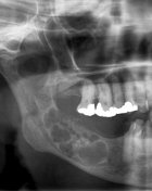
Image 8 CT (3D) ( update )

Image 9 CT (bone window) ( update )

Image 10 X-ray (OPG) ( update )

Image 11 CT (C+ arterial phase) ( create )


 Unable to process the form. Check for errors and try again.