
Anterior cerebral artery
Updates to Article Attributes
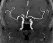
The anterior cerebral artery along with the middle cerebral artery, forms at the termination of the internal carotid artery. It is the smaller of the two, and arches antro-mediallyanteromedially to pass anterior to genu of the corpus callosum, dividing as it does so into its two major branches; pericallosal and callosomarginal arteries (see below).
It supplies the medial aspect of the cerebral hemispheres back to the parietal lobe.
Gross anatomy
Segments
The ACA is divided into three segments:
- A1 (horizontal): origin from the ICA to the anterior communicating artery (ACOM). ~14 mm in length
- A2 (vertical): from ACOM to the origin of the callosomarginal artery
- A3 (callosal): distal to the origin of the callosomarginal artery
Branches
There are two main branching patterns of the ACA. In the first the A2 gives off the callosomarginal artery (which lies in the cingulate sulcus), and continues as the pericallosal artery. In this configuration the terminal (cortical) branches are given off the callosomarginal artery.
In the second configuration the callosomarginal is absent and the terminal branches arise directly from the pericallosal.
-
A1:
medial lenticulostriate artery
recurrent artery of Heubner
anterior communicating artery
-
A2:
orbitofrontal artery
- frontopolar artery
- A3:
- pericallosal artery
callososeptal artery
Terminal (cortical) branches
Orbital branches, 2 or 3 in number, branch over the orbital surface of the frontal lobe supplying:
- olfactory cortex
- gyrus rectus
- medial orbital gyrus
They are named:
Frontal branches supply:
- corpus callosum (with the exception of the splenium)
- cingulate gyrus
- medial frontal gyrus
- paracentral lobule (as the branches reach over the vertex to supply a stip of cortex on the surface, they are responsible for supply to the lower limbs)
Parietal branches supply:
- precuneus
Central branches
Multiple small branches given off proximally (A1, ACOM, proximal A2) supply:
- anterior perforated substance
- lamina terminalis
- rostrum of the corpus callosum
- septum pellucidum
- anterior part of the putamen
- head of the caudate nucleus
- anteromedial part of the anterior limb of the internal capsule
The latter two (head of caudate and adjacent part of the internal capsule) are supplied by the recurrent artery of Heubner and associated smaller perforating branches. It has its origin near the A1-ACOM-A2 junction and can arise from all three, although usually it is from the A2. It curves back on itself and is at risk from ACOM aneurysm clipping.
Normal variantsVariant anatomy
- ACA fenestration with a reported incidence of 0-4% of A1 segment fenestration
- azygos ACA: ACA territories supplied by a single A2 trunk; incidence of ~2% (range 0.2-4.0%)
- ACA trifurcation: three A2 segments; incidence of ~7.5% (range 2-13%)
- bihemispheric ACA: hypoplastic A2 segment with contralateral A2 segment dominance supplying both ACA territories; incidence of ~4.5% (range 2-7%)
- A1 segment absence/hypoplasia, contralateral A1 segment dominance and supply to ipsilateral A2 segment by a large anterior communicating artery; 10 % of individuals demonstrate hypoplasia of A1 segment using a diameter 1.5 mm or smaller
- Asymmetry of A1 segment which is associated with ACA aneurysm
- persistent primitive olfactory artery 6
-<p>The<strong> anterior cerebral artery</strong> along with the <a href="/articles/middle-cerebral-artery">middle cerebral artery</a>, forms at the termination of the <a href="/articles/internal-carotid-artery-1">internal carotid artery</a>. It is the smaller of the two, and arches antro-medially to pass anterior to genu of the <a href="/articles/corpus-callosum">corpus callosum</a>, dividing as it does so into its two major branches; pericallosal and callosomarginal arteries (see below). </p><p>It supplies the medial aspect of the cerebral hemispheres back to the parietal lobe. </p><h4>Segments</h4><p>The ACA is divided into three segments:</p><ul>- +<p>The<strong> anterior cerebral artery</strong> along with the <a href="/articles/middle-cerebral-artery">middle cerebral artery</a>, forms at the termination of the <a href="/articles/internal-carotid-artery-1">internal carotid artery</a>. It is the smaller of the two, and arches anteromedially to pass anterior to genu of the <a href="/articles/corpus-callosum">corpus callosum</a>, dividing as it does so into its two major branches; pericallosal and callosomarginal arteries (see below). </p><p>It supplies the medial aspect of the cerebral hemispheres back to the parietal lobe. </p><h4>Gross anatomy</h4><h5>Segments</h5><p>The ACA is divided into three segments:</p><ul>
-</ul><h4>Branches</h4><p>There are two main branching patterns of the ACA. In the first the A2 gives off the callosomarginal artery (which lies in the cingulate sulcus), and continues as the pericallosal artery. In this configuration the terminal (cortical) branches are given off the callosomarginal artery.</p><p>In the second configuration the callosomarginal is absent and the terminal branches arise directly from the pericallosal.</p><ul>- +</ul><h5>Branches</h5><p>There are two main branching patterns of the ACA. In the first the A2 gives off the callosomarginal artery (which lies in the cingulate sulcus), and continues as the pericallosal artery. In this configuration the terminal (cortical) branches are given off the callosomarginal artery.</p><p>In the second configuration the callosomarginal is absent and the terminal branches arise directly from the pericallosal.</p><ul>
-</ul><h5>Terminal (cortical) branches</h5><p>Orbital branches, 2 or 3 in number, branch over the orbital surface of the frontal lobe supplying:</p><ul>- +</ul><h6>Terminal (cortical) branches</h6><p>Orbital branches, 2 or 3 in number, branch over the orbital surface of the frontal lobe supplying:</p><ul>
-</ul><p>Parietal branches supply:</p><ul><li>precuneus</li></ul><h5>Central branches</h5><p>Multiple small branches given off proximally (A1, ACOM, proximal A2) supply:</p><ul>- +</ul><p>Parietal branches supply:</p><ul><li>precuneus</li></ul><h6>Central branches</h6><p>Multiple small branches given off proximally (A1, ACOM, proximal A2) supply:</p><ul>
-</ul><p>The latter two (head of caudate and adjacent part of the internal capsule) are supplied by the <a href="/articles/recurrent_artery_of_huebner">recurrent artery of Heubner</a> and associated smaller perforating branches. It has its origin near the A1-ACOM-A2 junction and can arise from all three, although usually it is from the A2. It curves back on itself and is at risk from ACOM aneurysm clipping.</p><h4>Normal variants</h4><ul>- +</ul><p>The latter two (head of caudate and adjacent part of the internal capsule) are supplied by the <a href="/articles/recurrent_artery_of_huebner">recurrent artery of Heubner</a> and associated smaller perforating branches. It has its origin near the A1-ACOM-A2 junction and can arise from all three, although usually it is from the A2. It curves back on itself and is at risk from ACOM aneurysm clipping.</p><h4>Variant anatomy</h4><ul>
Image 2 Diagram ( update )

Image 3 DSA (angiography) ( update )

Image 4 Annotated image ( update )

Image 5 Diagram ( update )

Image 9 DSA (angiography) ( update )

Image 10 CT (C+ arterial phase) ( update )

Image 11 CT (C+ arterial phase) ( update )

Image 12 MRI ( update )


 Unable to process the form. Check for errors and try again.