
Cellulitis
Updates to Article Attributes
Cellulitis (more specifically referred to as superficial cellulitis) is an acute infection of the dermis and subcutaneous tissues. It results in pain, erythema, oedema, and warmth. Since the epidermis is not involved, cellulitis is not transmitted by person-to-person contact.
Pathology
Cellulitis occurs following disruption of the skin and invasion by microorganisms that may be indigenous flora, such as Staphylococcus aureus, or exogenous bacteria. Patients with peripheral vascular disease or diabetes are particularly susceptible to this type of infection, since minor injuries to the skin or cracked skin in the feet or toes can serve as a point of entry for infection.
Radiographic features
Ultrasound
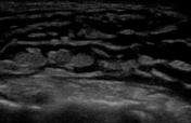
Ultrasound is usually the first investigation to evaluate a clinical suspicion of cellulitis. Normally the subcutaneous tissue is hypoechoic with few hyperechoic strands (representing connective tissue).
- In early cellulitis, there is a diffuse increase in the thickening and echogenicity of the subcutaneous tissue
. A helpful; helpful technique is to compare the tissue with contralateral side. - later, there is accumulation of fluid in the subcutaneous tissue, giving it a "cobble-stone appearance" (this appearance is not specific for cellulitis, but is seen in subcutaneous edema due to any cause)
.
CT
CT is used to accurately differentiate between superficial cellulitis and deep cellulitis (cellulitis associated with deep-seated infection).
In uncomplicated cellulitis, CT demonstrates skin thickening, septation of the subcutaneous fat, and thickening of the underlying superficial fascia. If the infection spreads to deeper tissues, deep cellulitis, soft-tissue abscess, infectious myositis, necrotizing fasciitis, and osteomyelitis can all be detected with CT.
Complications
Uncomplicated cellulitis is a clinical diagnosis and is treated conservatively with antibiotics and locally supportive measures. If the infection spreads to deeper tissues complications and associated pathology can occur, e.g. deep cellulitis, soft-tissue abscess, infectious myositis, necrotizing fasciitis, orosteomyelitis. Special consideration should be given to geriatric patients, in whom cellulitis of the lower extremities is more likely to develop intothrombophlebitis.
See also
-<li>In early cellulitis, there is a diffuse increase in the thickening and echogenicity of the subcutaneous tissue. A helpful technique is to compare the tissue with contralateral side. </li>-<li>later, there is accumulation of fluid in the subcutaneous tissue, giving it a "cobble-stone appearance" (this appearance is not specific for cellulitis, but is seen in subcutaneous edema due to any cause).</li>-</ul><h5>CT</h5><p>CT is used to accurately differentiate between superficial cellulitis and <a href="/articles/deep-cellulitis">deep cellulitis</a> (cellulitis associated with deep-seated infection). </p><p>In uncomplicated cellulitis, CT demonstrates skin thickening, septation of the <a href="/articles/subcutaneous-fat">subcutaneous fat</a>, and thickening of the underlying <a href="/articles/superficial-fascia">superficial fascia</a>. If the infection spreads to deeper tissues, deep cellulitis, soft-tissue abscess, infectious myositis, necrotizing fasciitis, and osteomyelitis can all be detected with CT.</p><h4>Complications</h4><p>Uncomplicated cellulitis is a clinical diagnosis and is treated conservatively with antibiotics and locally supportive measures. If the infection spreads to deeper tissues complications and associated pathology can occur, e.g. <a href="/articles/deep-cellulitis">deep cellulitis</a>, <a href="/articles/soft-tissue-abscess">soft-tissue abscess</a>, <a href="/articles/infectious-myositis">infectious myositis</a>, <a href="/articles/necrotizing-fasciitis">necrotizing fasciitis</a>, or <a href="/articles/osteomyelitis">osteomyelitis</a>. Special consideration should be given to geriatric patients, in whom cellulitis of the lower extremities is more likely to develop into <a href="/articles/thrombophlebitis">thrombophlebitis</a>.</p><h4>See also</h4><ul>- +<li>In early cellulitis, there is a diffuse increase in the thickening and echogenicity of the subcutaneous tissue; helpful technique is to compare the tissue with contralateral side</li>
- +<li>later, there is accumulation of fluid in the subcutaneous tissue, giving it a "cobble-stone appearance" (this appearance is not specific for cellulitis, but is seen in subcutaneous edema due to any cause)</li>
- +</ul><h5>CT</h5><p>CT is used to accurately differentiate between superficial cellulitis and <a href="/articles/deep-cellulitis">deep cellulitis</a> (cellulitis associated with deep-seated infection). </p><p>In uncomplicated cellulitis, CT demonstrates skin thickening, septation of the <a href="/articles/subcutaneous-fat">subcutaneous fat</a>, and thickening of the underlying <a href="/articles/superficial-fascia">superficial fascia</a>. If the infection spreads to deeper tissues, deep cellulitis, soft-tissue abscess, infectious myositis, necrotizing fasciitis, and osteomyelitis can all be detected with CT.</p><h4>Complications</h4><p>Uncomplicated cellulitis is a clinical diagnosis and is treated conservatively with antibiotics and locally supportive measures. If the infection spreads to deeper tissues complications and associated pathology can occur, e.g. <a href="/articles/deep-cellulitis">deep cellulitis</a>, <a href="/articles/soft-tissue-abscess">soft-tissue abscess</a>, <a href="/articles/infectious-myositis">infectious myositis</a>, <a href="/articles/necrotizing-fasciitis">necrotizing fasciitis</a>, or <a href="/articles/osteomyelitis">osteomyelitis</a>. Special consideration should be given to geriatric patients, in whom cellulitis of the lower extremities is more likely to develop into <a href="/articles/thrombophlebitis">thrombophlebitis</a>.</p><h4>See also</h4><ul>
-<li><a title="subcutaneous abscess" href="/articles/subcutaneous-abscess">subcutaneous abscess</a></li>- +<li><a href="/articles/subcutaneous-abscess">subcutaneous abscess</a></li>
Image 1 Photo ( create )

Image 3 CT (non-contrast) ( update )

Image 4 Ultrasound ( update )

Image 5 Ultrasound (Longitudinal) ( update )

Image 6 Ultrasound (Transverse) ( create )


 Unable to process the form. Check for errors and try again.