
Cerebral amyloid angiopathy
Updates to Article Attributes
Cerebral amyloid angiopathy (CAA) is a cerebrovascular disorder caused by the accumulation of cerebral amyloid-β (Aβ) in the tunica media and adventitia of leptomeningeal and cortical vessels of the brain. The resultant vascular fragility tends to manifest in normotensive elderly patients as lobar intracerebral haemorrhage. It is, along with Alzheimer disease, a common cerebral amyloid deposition disease.
Epidemiology
Cerebral amyloid angiopathy can be divided into sporadic (spontaneous) and familial forms.
Sporadic CAA
Cerebral amyloid angiopathy is a frequent incidental finding, found on screening gradient-recalled echo imaging in up to 16% of asymptomatic elderly patients 4. Autopsy studies have found a prevalence of approximately 5-9% in patients between 60 and 69 years, and 43-58% in patients over the age of 90 4.
Autopsies of patients who have evidence of Alzheimer disease have found cerebral amyloid angiopathy in the vast majority of cases (90%). This rate is still high (20-40%) in non-demented elderly individuals 14.
Importantly it is usually notassociated with systemic amyloidoses.
Familial CAA
Familial cerebral amyloid angiopathy describes a group of very rare disorders that are usually encountered as autosomal dominant conditions 14,21. Many of these disorders are only isolated to only a few families and they mainly differ from spontaneous CAA in an earlier age of onset, typically in middle to late middle age 14,21. Furthermore, they may also be part of multi-system or other central nervous system genetic disorders 14,21.
Examples of familial CAA include 21:
- Aß peptide with precursor protein APP (chromosome 21):
- CAA related to familial Alzheimer disease
- CAA in Down syndrome
- hereditary cerebral haemorrhage with amyloidosis (Dutch, Italian, Flemish, Iowa, Piedmont, Arctic types)
- ACys peptide with precursor protein cystatin C (chromosome 20): hereditary cerebral haemorrhage with amyloidosis Icelandic type
- ATTR peptide with precursor protein transthyretin (chromosome 18): meningovascular amyloidosis (see cerebral transthyretin-associated amyloidoses)
- AGel peptide with precursor protein gelsolin (chromosome 9): familial amyloidosis - Finnish type
- PrPSc peptide with precursor prion protein (chromosome 20): Gerstmann-Straussler-Scheinker disease
- ABri peptide with precursor protein ABri precursor protein (chromosome 13): familial British dementia (see case 17)
- ADan peptide with precursor protein ADan precursor protein (chromosome 13): familial Danish dementia
Clinical presentation
Manifestations of cortical vessel involvement:
- lobar haemorrhages or cerebellar haemorrhages: present as stroke, often with headache, focal neurological symptoms, seizures, and decreased conscious state 19
- cognitive impairment: occurs in three main patterns
- gradual decline: a vascular dementia thought to be secondary to lobar cerebral microhaemorrhages, ischaemic leukoencephalopathy, microinfarcts, and lobar lacunes 7,15, and occurs independently to cognitive impairment of Alzheimer disease 25
- step-wise decline: due to recurrent lobar haemorrhages 25
- rapidly-progressive decline: may be present in inflammatory cerebral amyloid angiopathy 25, which is discussed separately
The primary manifestation of leptomeningeal vessel involvement is due to convexity subarachnoid haemorrhage, which can present with transient focal neurological symptoms (TFNS) or "amyloid spells" 25. These TFNS are classically described as recurrent, stereotyped, spreading paraesthesias lasting several minutes but there is a wide spectrum of presentations encompassing both positive (spreading paraesthesia or visual symptoms) and negative (paresis, aphasia or dysphagia) phenomenology 17,25. These symptoms are most prominent with the convexity subarachnoid haemorrhage is localised to the central sulcus 16, which is in close proximity to the primary motor and sensory cortices 25.
Other manifestations of CAA, which are discussed separately, include:
- inflammatory cerebral amyloid angiopathy: an umbrella description for inflammatory reactions that present with rapidly-progressive cognitive decline, seizures, headache and stroke-like episodes (without haemorrhage) 1,11
- cerebral amyloidoma: mass-like lesions that have a varied presentation depending on the location of the amyloidoma
Pathology
Cerebral amyloid angiopathy is characterised by the deposition of amyloid in the tunica media and/or tunica adventitia of small and medium-sized arteries of the cerebral cortex and leptomeninges 4,20. This is associated with fibrinoid degeneration with separation of the tunica media and tunica intima, and microaneurysm formation 1.
There are a number of different proteins that can lead to intravascular amyloid deposition, however, the most common, as is the case in sporadic CAA, is Aß which is a short 42 amino acid peptide cleaved from amyloid precursor protein (APP) which is encoded on chromosome 21 20.
Aß is an eosinophilic, insoluble protein, located in the extracellular space. It stains with Congo red yielding classic apple-green birefringence when viewed with polarised light 3,20. When staining with thioflavin T and illuminated with ultraviolet light, the Aß deposits emit bright green fluorescence 20.
Associations
-
Alzheimer disease
- pathological cerebral amyloid angiopathy changes are seen in ~80% of those with Alzheimer disease (Aß-42) 5-13
- ~40% of those with cerebral amyloid angiopathy have Alzheimer dementia type symptoms
- Down syndrome 25
- chronic traumatic encephalopathy
- spongiform encephalitis
- other familial syndromes (as discussed above)
Radiographic features
Findings reflect the various manifestations of the disease:
- haemorrhage
-
intracerebral haemorrhage
- usually cortico-subcortical, in a so-called lobar location 22, but can also be seen in the cerebellum (especially in the cerebellar cortex or vermis) 24, may have finger-like projections 26
- tend to spare the basal ganglia and pons (cf. hypertensive 'deep' intracerebral haemorrhage)
- CT: initially hyperdense with hypodense perihaematomal oedema, often exerts positive mass-effect 25
- MRI: appearance will vary according to age of bleed (see blood on MRI) 25
- usually cortico-subcortical, in a so-called lobar location 22, but can also be seen in the cerebellum (especially in the cerebellar cortex or vermis) 24, may have finger-like projections 26
- cerebral microhaemorrhage
- defined as 2-10
millimetermillimetre, round or ovoid areas of haemorrhage, and tend to be corticosubcortical (grey-white matter junction) in distribution 25, but can also be in the superficial cerebellum 29- tend to spare the basal ganglia and pons (cf. hypertensive microhaemorrhages) 4,14
- CT: not appreciated 25
- MRI: only seen on T2* sequences (GRE, echo-planar, SWI) as regions of low-signal blooming artifact 12,25, not seen on conventional T1 and T2/FLAIR sequences 4
- defined as 2-10
-
convexity subarachnoid haemorrhage
- haemorrhage that is localised to one or more adjacent cortical sulci at the convexity of the brain
- tend to spare the basal cisterns, the Sylvian fissure, the interhemispheric fissure or the ventricles (cf. aneurysmal subarachnoid haemorrhage or perimesencephalic subarachnoid haemorrhage) 25
- CT: hyperdensity localised to one or more adjacent sulci, can be subtle 25
- MRI: appearance will vary according to the age of the bleed (see blood on MRI), but is best acutely seen on T2 FLAIR as a hyperintensity 18,25
- haemorrhage that is localised to one or more adjacent cortical sulci at the convexity of the brain
- cortical superficial siderosis
- thought to be a chronic sequela of convexity subarachnoid haemorrhage, including of haemorrhage that is asymptomatic 25
- not present infratentorially (cf. superficial siderosis of the CNS) 25
- CT: not appreciated 25
- MRI: curvilinear regions of signal drop-out localised to one or more sulci best seen on T2* sequences (GRE, echo-planar, SWI) 9,25
- thought to be a chronic sequela of convexity subarachnoid haemorrhage, including of haemorrhage that is asymptomatic 25
- cerebellar superficial siderosis 28
- similar to cortical superficial siderosis but involving the folia of the cerebellum, less prevalent than cortical superficial siderosis 28
- CT: not appreciated
- MRI: curvilinear regions of signal drop-out localised to one or more folia best seen on T2* sequences (GRE, echo-planar, SWI) 28
-
intracerebral haemorrhage
- ischaemia
- ischaemic leukoencephalopathy
- chronic lesions, indistinguishable from leukoaraiosis due to other aetiologies, but tends to have a periventricular and posterior predominance 25
- CT: diffuse hypodensity of the white matter 25
- MR: T2 hyperintensity of the white matter without involvement of subcortical U-fibres (cf. cerebral amyloid angiopathy related inflammation) 7,25
- microinfarcts and lobar lacunes
- acute cortico-subcortical lesions; lobar lacunes are 3-15 millimetres in size while microinfarcts are smaller 25
- CT: not appreciated 25
- MRI: same signal changes as in acute ischaemic stroke, most pronounced on DWI 25
- ischaemic leukoencephalopathy
- others
- dilated perivascular spaces of the centrum semiovale
- dilation of normal perivascular spaces in the centrum semiovale 25
- tend to spare the basal ganglia and pons (cf. hypertensive dilated perivascular spaces) 25
- CT: not appreciated 25
- MRI: best appreciated on T2 images as CSF-signal structures with a varied appearance depending on the orientation of their draining vessel 25
- dilation of normal perivascular spaces in the centrum semiovale 25
- cortical atrophy
- CT: not appreciated 25
- MRI: not readily appreciated on conventional sequences, requires cortical surface reconstructions 25
- dilated perivascular spaces of the centrum semiovale
Radiographic features of inflammatory cerebral amyloid angiopathy and cerebral amyloidoma are discussed separately.
Diagnostic criteria
The Boston criteria 7 and newer Modified Boston criteria 9 are a combination of clinical, radiographic and pathological criteria which are used to assess the probability of cerebral amyloid angiopathy. These criteria require patients to have either biopsy specimens and/or brain MRI data available 7,9. Additionally, the Edinburgh criteria for lobar intracerebral haemorrhage associated with cerebral amyloid angiopathy can be utilised, especially for patients with a lobar intracerebral haemorrhage without an MRI 26.
Treatment and prognosis
There is currently no disease-modifying treatment available 27. Additionally, there are no guidelines regarding use of antiplatelet, anticoagulant, or thrombolytic drugs in patients with CAA, all medications which have been shown to increase the risk of disabling haemorrhage in this patient group 27.
Differential diagnosis
Radiological differential diagnosis, particularly of cerebral microhaemorrhages, includes:
-
hypertensive microangiopathy
- haemorrhages, including microhaemorrhages, are typically located in basal ganglia, pons and cerebellum
- not associated with subarachnoid haemorrhage or superficial siderosis
-
multiple cavernoma syndrome
- lesions have a random distribution
- random size, although Zabramski classification type IV cavernous malformations are indistinguishable from cerebral microhaemorrhages related to CAA
- often characteristic cavernous malformations can be identified
-
haemorrhagic metastases (e.g. melanoma)
- lesions have a variable size and can often be larger than microhaemorrhages
- enhancing
-
diffuse axonal injury
- lesions are typically located at the grey-white matter junction, in the corpus callosum and in more severe cases, in the brainstem
-
neurocysticercosis
- nodular calcified stage visible on CT or phase-filtered SWI
- random distribution
-
fat embolism syndrome
- 'starfield' pattern of distribution
- lesions also show restricted diffusion on DWI and are likely visible on other sequences
-
radiation-induced vasculopathy
- microhaemorrhages have a very similar appearance (similar pathophysiology)
- distribution related to the treatment field
-<li>defined as 2-10 millimeter, round or ovoid areas of haemorrhage, and tend to be corticosubcortical (grey-white matter junction) in distribution <sup>25</sup>, but can also be in the superficial cerebellum <sup>29</sup><ul><li>tend to spare the <a href="/articles/basal-ganglia">basal ganglia</a> and <a href="/articles/pons">pons</a> (cf. <a href="/articles/hypertensive-microangiopathy">hypertensive microhaemorrhages</a>) <sup>4,14</sup>- +<li>defined as 2-10 millimetre, round or ovoid areas of haemorrhage, and tend to be corticosubcortical (grey-white matter junction) in distribution <sup>25</sup>, but can also be in the superficial cerebellum <sup>29</sup><ul><li>tend to spare the <a href="/articles/basal-ganglia">basal ganglia</a> and <a href="/articles/pons">pons</a> (cf. <a href="/articles/hypertensive-microangiopathy">hypertensive microhaemorrhages</a>) <sup>4,14</sup>
Image 3 Diagram ( create )
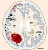
Image 5 MRI (SWI) ( update )

Image 6 MRI (SWI) ( update )

Image 7 CT (non-contrast) ( update )

Image 8 MRI (Gradient Echo) ( update )

Image 9 MRI ( update )

Image 10 CT (non-contrast) ( update )

Image 11 MRI (T1) ( update )

Image 12 MRI (T2) ( update )

Image 13 MRI (Gradient Echo) ( update )

Image 14 MRI (SWI) ( update )

Image 15 MRI (Gradient Echo) ( update )

Image 16 MRI (SWI) ( update )

Image 17 MRI (Gradient Echo) ( update )

Image 18 MRI (Gradient Echo) ( update )

Image 19 MRI (SWI MIP) ( update )

Image 20 MRI (Gradient Echo) ( update )

Image 22 MRI (SWI) ( update )

Image 23 MRI (SWI MIP) ( update )

Image 24 MRI (Gradient Echo) ( update )


 Unable to process the form. Check for errors and try again.