
Cervical spine series
Updates to Article Attributes
Body
was changed:
The cervical spine series is a set of radiographs taken to investigate the bony structures of the cervical spine, albeit commonly replaced by the CT, the cervical spine series is an essential trauma radiograph for all radiographers to understand.
Indications
Cervical spine radiographs are indicated for a variety of settings including 1-3:
- trauma
- infection
- atypical pain
- limb pain
- osteoporosis
- degenerative changes
A decision to pursue c-spine imaging of any kind should be cross-referenced with the 'Canadian C-Spine Rule' for c-spine imaging due to its high sensitivity and specificity 4
Projections
Standard projections
Note:in the absence of CT 5 views of the C-spine should be performed, AP, lateral, obliques and odontoid 5 .
-
AP
- anteroposterior projection of the cervical spine demonstrating the vertebral bodies and intervertebral spaces
-
lateral
- often utilised in trauma demonstrated
- zygapophyseal joints
- soft tissue structures around the c spine
- spinous processes
- anterior-posterior relationship of the vertebral bodies
- often utilised in trauma demonstrated
-
odontoid
- also known as a 'peg' projection it demonstrates the C1 (atlas) and C2 (axis)
Additional projections
-
AP oblique
- demonstrates the intervertebral foramina of the side positioned further from the image receptor
-
PA oblique
- demonstrated the intervertebral foramina of the side positioned closer to the image receptor
Additional projections
-
cervicothoracic view
- modified lateral projection of the cervical spine to visualise the C7/T1 junction
-
flexion-extension lateral
- specialised projections of the cervical spine often requested to assess for spinal stability.
-
Fuchs view
- nonangled AP radiograph of C1 and C2. it should not be used in a trauma setting
-<li>trauma </li>- +<li>trauma</li>
-</ul><p>A decision to pursue c-spine imaging of any kind should be cross-referenced with the 'Canadian C-Spine Rule' for c-spine imaging due to its high sensitivity and specificity <sup>4</sup></p><h4>Projections</h4><h5>Standard projections</h5><p>Note: in the absence of CT 5 views of the C-spine should be performed, AP, lateral, obliques and odontoid <sup>5</sup> .</p><ul>- +</ul><p>A decision to pursue c-spine imaging of any kind should be cross-referenced with the 'Canadian C-Spine Rule' for c-spine imaging due to its high sensitivity and specificity <sup>4</sup></p><h4>Projections</h4><h5>Standard projections</h5><p><strong>Note: </strong>in the absence of CT 5 views of the C-spine should be performed, AP, lateral, obliques and odontoid <sup>5</sup> .</p><ul>
-<a title="Cervical spine (AP view)" href="/articles/cervical-spine-ap-view">AP</a> <ul><li>anteroposterior projection of the cervical spine demonstrating the vertebral bodies and intervertebral spaces</li></ul>- +<a href="/articles/cervical-spine-ap-view">AP</a> <ul><li>anteroposterior projection of the cervical spine demonstrating the vertebral bodies and intervertebral spaces</li></ul>
-<a title="Cervical spine (lateral view)" href="/articles/cervical-spine-lateral-view">lateral </a> <ul><li>often utilised in trauma demonstrated<ul>- +<a href="/articles/cervical-spine-lateral-view">lateral </a> <ul><li>often utilised in trauma demonstrated<ul>
-<a title="Cervical spine (odontoid view)" href="/articles/cervical-spine-odontoid-view">odontoid </a> <ul><li>also known as a 'peg' projection it demonstrates the <a href="/articles/atlas-c1">C1 (atlas)</a> and <a href="/articles/axis-c2">C2 (axis)</a>- +<a href="/articles/cervical-spine-odontoid-view">odontoid </a> <ul><li>also known as a 'peg' projection it demonstrates the <a href="/articles/atlas-c1">C1 (atlas)</a> and <a href="/articles/axis-c2">C2 (axis)</a>
-</ul><h5>Additional projections</h5><ul>-<a title="Cervical spine (AP oblique view)" href="/articles/cervical-spine-ap-oblique-view">AP oblique </a><ul><li>demonstrates the intervertebral foramina of the side positioned further from the image receptor</li></ul>- +<a href="/articles/cervical-spine-ap-oblique-view">AP oblique</a><ul><li>demonstrates the intervertebral foramina of the side positioned further from the image receptor</li></ul>
-<a title="Cervical spine (PA oblique view)" href="/articles/cervical-spine-pa-oblique-view">PA oblique </a><ul><li>demonstrated the intervertebral foramina of the side positioned closer to the image receptor </li></ul>- +<a href="/articles/cervical-spine-pa-oblique-view">PA oblique</a><ul><li>demonstrated the intervertebral foramina of the side positioned closer to the image receptor</li></ul>
- +</ul><h5>Additional projections</h5><ul>
-<a title="Cervical spine (swimmer's lateral view)" href="/articles/cervical-spine-swimmers-lateral-view-1">cervicothoracic view</a><ul><li>modified lateral projection of the cervical spine to visualise the C7/T1 junction</li></ul>- +<a href="/articles/cervical-spine-swimmers-lateral-view-1">cervicothoracic view</a><ul><li>modified lateral projection of the cervical spine to visualise the C7/T1 junction</li></ul>
-<a title="Cervical spine (flexion-extension views)" href="/articles/cervical-spine-flexion-extension-views-1">flexion-extension lateral </a><ul><li>specialised projections of the cervical spine often requested to assess for spinal stability.</li></ul>- +<a href="/articles/cervical-spine-flexion-extension-views-1">flexion-extension lateral </a><ul><li>specialised projections of the cervical spine often requested to assess for spinal stability.</li></ul>
-<a title="Cervical spine (Fuchs view)" href="/articles/cervical-spine-fuchs-view">Fuchs view</a><ul><li>nonangled AP radiograph of C1 and C2. it should not be used in a trauma setting</li></ul>- +<a href="/articles/cervical-spine-fuchs-view">Fuchs view</a><ul><li>nonangled AP radiograph of C1 and C2. it should not be used in a trauma setting</li></ul>
References changed:
- 1. Houben PH, der van Weijden T, Sijbrandij J, Grol RP, Winkens RA. Reasons for ordering spinal x-ray investigations: how they influence general practitioners' management. (2006) Canadian family physician Medecin de famille canadien. 52 (10): 1266-7. <a href="https://www.ncbi.nlm.nih.gov/pubmed/17279187">Pubmed</a> <span class="ref_v4"></span>
- 2. John Lampignano, Leslie E. Kendrick. Bontrager's Textbook of Radiographic Positioning and Related Anatomy. (2017) ISBN: 9780323399661
- 3. Thompson WL, Stiell IG, Clement CM et-al. Association of injury mechanism with the risk of cervical spine fractures. CJEM. 2009;11 (1): 14-22. Pubmed citation
- 4. Saragiotto BT, Michaleff ZA. The Canadian C-Spine Rule. (2016) Journal of physiotherapy. 62 (3): 170. <a href="https://doi.org/10.1016/j.jphys.2016.04.001">doi:10.1016/j.jphys.2016.04.001</a> - <a href="https://www.ncbi.nlm.nih.gov/pubmed/27161303">Pubmed</a> <span class="ref_v4"></span>
- 5. Goergen S, D Varma and H Ackland et al. Adult Cervical Spine Trauma. Education Modules for Appropriate Imaging Referrals: Royal Australian and New Zealand College of Radiologists, 2015. <a href="https://www.ranzcr.com/documents/3831-print-version-adult-c-spine/file" target="_blank">[Link]</a>.
Sections changed:
- Radiography
Systems changed:
- Spine
Images Changes:
Image 2 X-ray (Frontal) ( create )

Image 3 X-ray (Peg view) ( create )

Image 4 X-ray (Oblique) ( create )

Image 5 X-ray (Oblique) ( create )

Image 6 Annotated image ( create )
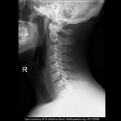
Image 7 X-ray (Swimmers lateral) ( create )

Image 8 X-ray (Fuchs view) ( create )

Image 9 X-ray (Lateral) ( create )

Image 10 X-ray (Lateral) ( create )


 Unable to process the form. Check for errors and try again.