
Cleft lip and palate
Updates to Article Attributes
Cleft lip and palate is one of the commonest neonatal facial anomalies. In ~80% of cases, the two features tend to occur together 6.
Epidemiology
It is estimated to occur with an incidence of ~1 in 700-1,000-1000 live births 1. This can increase to 4% for a sibling of a previously affected fetus and up to 10% for a sibling of two previously affected infants 9. There
There is a recognised racial predilection. For example, the United States, has one of the highest rates in terms of prevalence in Native Americans (~4 in 1,0001000), followed by Asian (1.5-2.0 in 1,0001000), CaucasianWhite (1 in 1,0001000), and African AmericanBlack (0.3 in 1,0001000) populations.
Associations
Other associated anomalies can occur in up to 30-50% of cases 3,6. They are protean and include:
-
aneuploidic syndromic: tends to occur with types II, III and IV 5
-
non-aneuploidic syndromic
ectrodactyly-ectodermal dysplasia-clefting syndrome-EEC syndrome
Majewski syndrome: short rib-polydactyly syndrome type II
oro-facial-digital syndrome type 1 (OFDS type 1): particularly tends to give a median cleft
certain types of short rib-polydactyly syndromes, e.g. type IV
-
exposure to in utero substances
-
non-aneuploidic non-syndromic
holoprosencephaly: often with type IV
Pathology
The lip forms between the 4th andto 7th weeks of pregnancy. The roof of the mouth (palate) is formed between the 6th andto 9th weeks of pregnancy. The condition results during the 4th to 6th weeks of gestation from a failure of fusion of one or both of the medial nasal prominences. These initially occur as paired medial nasal processes and failure of fusion with each other or with the maxillary processes will result in cleft lip with or without a cleft palate. Clefts in the anterior palate disrupt this normal line of fusion, resulting in the discontinuity in the smooth, C-shape contour of the alveolar ridge at the junction between the lateral incisors and canine tooth sockets, extending posteriorly to the incisive foramen. Much less commonly, the defect may occur between the medial and lateral incisors.
In a bilateral cleft lip and palate, there is a premaxillary protrusion that is typically seen as a paranasal echogenic mass. The premaxillary protrusion results from instability of the facial structures during embryologic development, which produces uninhibited growth of the vomer and premaxillary bones and soft tissues. Although this occurs with a bilateral complete cleft lip and palate, it does not occur with other types of facial clefts or with cleft palate alone.
Genetics
In a sizeable proportion, it is sporadic, although various inheritance patterns have been described 10.
LateralityLocation
The majority are unilateral and 70% of these are on the left.
Classification
Several classification systems exist. The Nyberg 1995 antenatal ultrasound classification system is one that correlates very well with the severity of the defect with outcomes and is divided into five types 5:
type I: isolated cleft lip alone
type II: unilateral cleft lip and palate
type III: bilateral cleft lip and palate
type IV: midline/median cleft lip and palate
type V: facial clefts associated with the amniotic band syndrome or the limb-body-wall complex
An isolated cleft palate is almost impossible to diagnose in utero and is not part of this classification.
Genetics
In a sizeable proportion, it is sporadic, although various inheritance patterns have been described 10.
Radiographic features
UltrasoundAntenatal ultrasound
Sonographic features can be variable depending on the exact type of cleft anomaly. In general, an upper lip defect may be seen and is best appreciated on angled coronal scanning. A vertical hypoechoic region through the fetal upper lip usually represents the defect in the cleft lip. This finding may be corroborated by a similar defect of the soft tissues of the upper lip overlying the maxilla in the axial plane.
The palate can be examined in the transverse (axial) plane. 3D ultrasound may further assist in diagnosis. It is good practice to comment on fetal swallowing in real time as the ultrasound is performed.
Type-specific sonographic features include:
type III: may be seen as a premaxillary echogenic mass 8
Ancillary sonographic features
there can be polyhydramnios if the defect is severe and impairs swallowing
Treatment and prognosis
The overall prognosis is variable depending on associated anomalies with isolated lateral clefts (types I and II) having the best prognosis.
See also
-<p><strong>Cleft lip</strong> <strong>and palate</strong> is one of the commonest neonatal facial anomalies. In ~80% of cases, the two features tend to occur together <sup>6</sup>.</p><h4>Epidemiology</h4><p>It is estimated to occur with an incidence of ~1 in 700-1,000 live births <sup>1</sup>. This can increase to 4% for a sibling of a previously affected fetus and up to 10% for a sibling of two previously affected infants <sup>9</sup>. There is a recognised racial predilection. For example, the United States, has one of the highest rates in terms of prevalence in Native Americans (~4 in 1,000), followed by Asian (1.5-2.0 in 1,000), Caucasian (1 in 1,000), and African American (0.3 in 1,000) populations.</p><h5>Associations</h5><p>Other associated anomalies can occur in up to 30-50% of cases <sup>3,6</sup>. They are <a href="/articles/protean">protean</a> and include</p><ul>- +<p><strong>Cleft lip</strong> <strong>and palate</strong> is one of the commonest neonatal facial anomalies. In ~80% of cases, the two features tend to occur together <sup>6</sup>.</p><h4>Epidemiology</h4><p>It is estimated to occur with an incidence of ~1 in 700-1000 live births <sup>1</sup>. This can increase to 4% for a sibling of a previously affected fetus and up to 10% for a sibling of two previously affected infants <sup>9</sup>. </p><p>There is a recognised racial predilection. For example, the United States has one of the highest rates in terms of prevalence in Native Americans (~4 in 1000), followed by Asian (1.5-2 in 1000), White (1 in 1000), and Black (0.3 in 1000) populations.</p><h5>Associations</h5><p>Other associated anomalies can occur in up to 30-50% of cases <sup>3,6</sup>. They are <a href="/articles/protean">protean</a> and include:</p><ul>
-<strong>aneuploidic syndromic:</strong> tends to occur with types II, III and IV <sup>5</sup><ul>-<li><a href="/articles/patau-syndrome">trisomy 13</a></li>-<li><a href="/articles/edwards-syndrome-1">trisomy 18</a></li>- +<p><strong>aneuploidic syndromic:</strong> tends to occur with types II, III and IV <sup>5</sup></p>
- +<ul>
- +<li><p><a href="/articles/patau-syndrome">trisomy 13</a></p></li>
- +<li><p><a href="/articles/edwards-syndrome-1">trisomy 18</a></p></li>
-<strong>non-aneuploidic syndromic</strong><ul>-<li>-<a href="/articles/ectrodactyly-ectodermal-dysplasia-facial-clefting-syndrome">ectrodactyly-ectodermal dysplasia-clefting syndrome</a>-<a href="/articles/eec-syndrome">EEC syndrome</a>-</li>-<li><a href="/articles/frontonasal-dysplasia">frontonasal dysplasia</a></li>-<li><a href="/articles/fryns-syndrome-1">Fryns syndrome</a></li>-<li><a href="/articles/gorlin-goltz-syndrome-1">Gorlin syndrome</a></li>-<li><a href="/articles/juberg-hayward-syndrome-1">Juberg-Hayward syndrome</a></li>-<li><a href="/articles/kallmann-syndrome">Kallmann syndrome</a></li>-<li>-<a href="/articles/majewski-syndrome">Majewski syndrome</a>: short rib-polydactyly syndrome type II</li>-<li>-<a href="/articles/nager-syndrome">Nager syndrome</a> <sup>10</sup>-</li>-<li>-<a href="/articles/oro-facial-digital-syndrome-type-1-ofds-type-1">oro-facial-digital syndrome type 1 (OFDS type 1)</a>: particularly tends to give a median cleft</li>-<li><a href="/articles/roberts-syndrome">Roberts syndrome</a></li>-<li>certain types of <a href="/articles/short-rib-polydactyly-syndrome">short rib-polydactyly syndromes</a>, e.g. type IV</li>-<li><a href="/articles/stickler-syndrome">Stickler syndrome</a></li>-<li><a href="/articles/thrombocytopenia-with-absent-radius-syndrome-1">TAR syndrome</a></li>-<li><a href="/articles/vacterl-association-1">VACTERL association</a></li>-<li>-<a href="/articles/van-der-woude-syndrome-vws">van der Woude syndrome (VWS)</a> <sup>7</sup>-</li>-<li>exposure to in utero substances<ul>-<li><a href="/articles/fetal-hydantoin-syndrome">fetal hydantoin syndrome</a></li>-<li><a href="/articles/fetal-valproate-syndrome">fetal valproate syndrome</a></li>- +<p><strong>non-aneuploidic syndromic</strong></p>
- +<ul>
- +<li><p><a href="/articles/ectrodactyly-ectodermal-dysplasia-facial-clefting-syndrome">ectrodactyly-ectodermal dysplasia-clefting syndrome</a>-<a href="/articles/eec-syndrome">EEC syndrome</a></p></li>
- +<li><p><a href="/articles/frontonasal-dysplasia">frontonasal dysplasia</a></p></li>
- +<li><p><a href="/articles/fryns-syndrome-1">Fryns syndrome</a></p></li>
- +<li><p><a href="/articles/gorlin-goltz-syndrome-1">Gorlin syndrome</a></p></li>
- +<li><p><a href="/articles/juberg-hayward-syndrome-1">Juberg-Hayward syndrome</a></p></li>
- +<li><p><a href="/articles/kallmann-syndrome">Kallmann syndrome</a></p></li>
- +<li><p><a href="/articles/majewski-syndrome">Majewski syndrome</a>: short rib-polydactyly syndrome type II</p></li>
- +<li><p><a href="/articles/nager-syndrome">Nager syndrome</a> <sup>10</sup></p></li>
- +<li><p><a href="/articles/oro-facial-digital-syndrome-type-1-ofds-type-1">oro-facial-digital syndrome type 1 (OFDS type 1)</a>: particularly tends to give a median cleft</p></li>
- +<li><p><a href="/articles/roberts-syndrome">Roberts syndrome</a></p></li>
- +<li><p>certain types of <a href="/articles/short-rib-polydactyly-syndrome">short rib-polydactyly syndromes</a>, e.g. type IV</p></li>
- +<li><p><a href="/articles/stickler-syndrome">Stickler syndrome</a></p></li>
- +<li><p><a href="/articles/thrombocytopenia-with-absent-radius-syndrome-1">TAR syndrome</a></p></li>
- +<li><p><a href="/articles/vacterl-association-1">VACTERL association</a></p></li>
- +<li><p><a href="/articles/van-der-woude-syndrome-vws">van der Woude syndrome (VWS)</a> <sup>7</sup></p></li>
- +<li>
- +<p>exposure to in utero substances</p>
- +<ul>
- +<li><p><a href="/articles/fetal-hydantoin-syndrome">fetal hydantoin syndrome</a></p></li>
- +<li><p><a href="/articles/fetal-valproate-syndrome">fetal valproate syndrome</a></p></li>
-<strong>non-aneuploidic non-syndromic</strong><ul>-<li><a href="/articles/dandy-walker-malformation-1">Dandy-Walker continuum</a></li>-<li>-<a href="/articles/holoprosencephaly">holoprosencephaly</a>: often with type IV</li>-<li><a href="/articles/pentalogy-of-cantrell-3">pentalogy of Cantrell</a></li>- +<p><strong>non-aneuploidic non-syndromic</strong></p>
- +<ul>
- +<li><p><a href="/articles/dandy-walker-malformation-1">Dandy-Walker continuum</a></p></li>
- +<li><p><a href="/articles/holoprosencephaly">holoprosencephaly</a>: often with type IV</p></li>
- +<li><p><a href="/articles/pentalogy-of-cantrell-3">pentalogy of Cantrell</a></p></li>
-</ul><h4>Pathology</h4><p>The lip forms between the 4<sup>th</sup> and 7<sup>th</sup> weeks of pregnancy. The roof of the mouth (<a href="/articles/palate">palate</a>) is formed between the 6<sup>th </sup>and 9<sup>th</sup> weeks of pregnancy. The condition results during the 4<sup>th</sup> to 6<sup>th</sup> weeks of gestation from a failure of fusion of one or both of the <a href="/articles/medial-nasal-prominences">medial nasal prominences</a>. These initially occur as paired medial nasal processes and failure of fusion with each other or with the maxillary processes will result in cleft lip with or without a cleft palate. Clefts in the anterior palate disrupt this normal line of fusion, resulting in the discontinuity in the smooth, C-shape contour of the alveolar ridge at the junction between the lateral incisors and canine tooth sockets, extending posteriorly to the <a href="/articles/incisive-foramen">incisive foramen</a>. Much less commonly, the defect may occur between the medial and lateral incisors. </p><p>In a bilateral cleft lip and palate, there is a <a href="/articles/premaxillary-protrusion">premaxillary protrusion</a> that is typically seen as a paranasal echogenic mass. The premaxillary protrusion results from instability of the facial structures during embryologic development, which produces uninhibited growth of the <a href="/articles/vomer">vomer</a> and premaxillary bones and soft tissues. Although this occurs with a bilateral complete cleft lip and palate, it does not occur with other types of facial clefts or with cleft palate alone.</p><h5>Genetics</h5><p>In a sizeable proportion, it is sporadic, although various inheritance patterns have been described <sup>10</sup>.</p><h5>Laterality</h5><p>The majority are unilateral and 70% of these are on the left<strong>.</strong></p><h5>Classification</h5><p>Several classification systems exist. The <a href="/articles/nyberg-1995-antenatal-ultrasound-classification-system">Nyberg 1995 antenatal ultrasound classification system</a> is one that correlates very well with the severity of the defect with outcomes and is divided into five types <sup>5</sup>:</p><ul>-<li>-<strong>type I:</strong> isolated cleft lip alone </li>-<li>-<strong>type II:</strong> unilateral cleft lip and palate </li>-<li>-<strong>type III:</strong> bilateral cleft lip and palate</li>-<li>-<strong>type IV:</strong> midline/median cleft lip and palate </li>-<li>-<strong>type V:</strong> <a href="/articles/facial-clefts">facial clefts</a> associated with the <a href="/articles/amniotic-band-syndrome">amniotic band syndrome</a> or the <a href="/articles/limb-body-wall-complex">limb-body-wall complex</a>-</li>-</ul><p>An <a href="/articles/isolated-cleft-palate">isolated cleft palate</a> is almost impossible to diagnose in utero and is not part of this classification.</p><h4>Radiographic features</h4><h5>Ultrasound</h5><p>Sonographic features can be variable depending on the exact type of cleft anomaly. In general, an upper lip defect may be seen and is best appreciated on angled coronal scanning. A vertical hypoechoic region through the fetal upper lip usually represents the defect in cleft lip. This finding may be corroborated by a similar defect of the soft tissues of the upper lip overlying the <a href="/articles/maxilla">maxilla</a> in the axial plane.</p><p>The palate can be examined in the transverse (axial) plane. <a href="/articles/3d-ultrasound">3D ultrasound</a> may further assist in diagnosis. It is good practice to comment on fetal swallowing in real time as the ultrasound is performed.</p><p>Type-specific sonographic features include:</p><ul><li>type III: may be seen as a premaxillary echogenic mass <sup>8</sup>-</li></ul><h6>Ancillary sonographic features</h6><ul><li>there can be <a href="/articles/polyhydramnios">polyhydramnios</a> if the defect is severe and impairs swallowing</li></ul><h4>Treatment and prognosis</h4><p>The overall prognosis is variable depending on associated anomalies with isolated lateral clefts (types I and II) having the best prognosis.</p><h4>See also</h4><ul>-<li><a href="/articles/isolated-cleft-palate">isolated cleft palate</a></li>-<li><a href="/articles/facial-clefts">facial clefts</a></li>-<li><a href="/articles/mesiodens">mesiodens</a></li>- +</ul><h4>Pathology</h4><p>The lip forms between the 4<sup>th</sup> to 7<sup>th</sup> weeks of pregnancy. The roof of the mouth (<a href="/articles/palate">palate</a>) is formed between the 6<sup>th </sup>to 9<sup>th</sup> weeks of pregnancy. The condition results during the 4<sup>th</sup> to 6<sup>th</sup> weeks of gestation from a failure of fusion of one or both of the <a href="/articles/medial-nasal-prominences">medial nasal prominences</a>. These initially occur as paired medial nasal processes and failure of fusion with each other or with the maxillary processes will result in cleft lip with or without a cleft palate. Clefts in the anterior palate disrupt this normal line of fusion, resulting in the discontinuity in the smooth, C-shape contour of the alveolar ridge at the junction between the lateral incisors and canine tooth sockets, extending posteriorly to the <a href="/articles/incisive-foramen">incisive foramen</a>. Much less commonly, the defect may occur between the medial and lateral incisors. </p><p>In a bilateral cleft lip and palate, there is a <a href="/articles/premaxillary-protrusion">premaxillary protrusion</a> that is typically seen as a paranasal echogenic mass. The premaxillary protrusion results from instability of the facial structures during embryologic development, which produces uninhibited growth of the <a href="/articles/vomer">vomer</a> and premaxillary bones and soft tissues. Although this occurs with a bilateral complete cleft lip and palate, it does not occur with other types of facial clefts or with cleft palate alone.</p><h5>Location</h5><p>The majority are unilateral and 70% of these are on the left<strong>.</strong></p><h5>Classification</h5><p>Several classification systems exist. The <a href="/articles/nyberg-1995-antenatal-ultrasound-classification-system">Nyberg 1995 antenatal ultrasound classification system</a> is one that correlates very well with the severity of the defect with outcomes and is divided into five types <sup>5</sup>:</p><ul>
- +<li><p><strong>type I:</strong> isolated cleft lip alone </p></li>
- +<li><p><strong>type II:</strong> unilateral cleft lip and palate </p></li>
- +<li><p><strong>type III:</strong> bilateral cleft lip and palate</p></li>
- +<li><p><strong>type IV:</strong> midline/median cleft lip and palate </p></li>
- +<li><p><strong>type V:</strong> <a href="/articles/facial-clefts">facial clefts</a> associated with the <a href="/articles/amniotic-band-syndrome">amniotic band syndrome</a> or the <a href="/articles/limb-body-wall-complex">limb-body-wall complex</a></p></li>
- +</ul><p>An <a href="/articles/isolated-cleft-palate">isolated cleft palate</a> is almost impossible to diagnose in utero and is not part of this classification.</p><h5>Genetics</h5><p>In a sizeable proportion, it is sporadic, although various inheritance patterns have been described <sup>10</sup>.</p><h4>Radiographic features</h4><h5>Antenatal ultrasound</h5><p>Sonographic features can be variable depending on the exact type of cleft anomaly. In general, an upper lip defect may be seen and is best appreciated on angled coronal scanning. A vertical hypoechoic region through the fetal upper lip usually represents the defect in the cleft lip. This finding may be corroborated by a similar defect of the soft tissues of the upper lip overlying the <a href="/articles/maxilla">maxilla</a> in the axial plane.</p><p>The palate can be examined in the transverse (axial) plane. <a href="/articles/3d-ultrasound">3D ultrasound</a> may further assist in diagnosis. It is good practice to comment on fetal swallowing in real time as the ultrasound is performed.</p><p>Type-specific sonographic features include:</p><ul><li><p>type III: may be seen as a premaxillary echogenic mass <sup>8</sup></p></li></ul><h6>Ancillary sonographic features</h6><ul><li><p>there can be <a href="/articles/polyhydramnios">polyhydramnios</a> if the defect is severe and impairs swallowing</p></li></ul><h4>Treatment and prognosis</h4><p>The overall prognosis is variable depending on associated anomalies with isolated lateral clefts (types I and II) having the best prognosis.</p><h4>See also</h4><ul>
- +<li><p><a href="/articles/isolated-cleft-palate">isolated cleft palate</a></p></li>
- +<li><p><a href="/articles/facial-clefts">facial clefts</a></p></li>
- +<li><p><a href="/articles/mesiodens">mesiodens</a></p></li>
Image 1 Photo ( update )
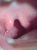
Image 2 Ultrasound ( update )

Image 3 Ultrasound ( update )

Image 4 Ultrasound (Transverse) ( update )

Image 5 Ultrasound (3d) ( update )
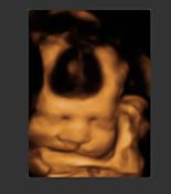
Image 8 Ultrasound ( update )

Image 9 Ultrasound ( update )


 Unable to process the form. Check for errors and try again.