
Dual-energy CT (clinical applications)
Updates to Article Attributes
Dual energy-energy CT is becoming increasing more common in clinical practice due to the rapid rise in computer technology and the expanding literature exhibiting widevast advantages over conventional single energy CT.
Virtual non-contrast
There is a potential to eliminate the need for pre-contrast imaging, using complex subtraction algorithms based on the two datasets known as virtual unenhanced imaging 1.
The acquired images are automatically reconstructed to three separate image sets: 80 kVp, 140 kVp and mixed 80:140 kVp image with the weighting factor of 0.4 (40% image information from the 80 kVp image and 60% information from the 140 kVp image). The weighting factors can be adjusted, to achieve the desirable effect. The 80 kvP images have higher contrast attenuation but intrinsically lower SNRsignal to noise ratio and smaller FOVfield of view. The 140 kVp images have lowerless contrast attenuation better SNRsignal to noise ratio and full FOVfield of view.
Material decomposition can be further performed on a dedicated workstation to create different image setting including iodine map (virtual contrast image), iodine subtraction (virtual non-contrast image) and bone mask (bone and calcium subtraction). A further perfusion blood volume colour coded images can be created by using a grey or colour scale. This perfusion blood volume images reflect the lung perfusion at a single time point. Thus they are only a surrogate perfusion images.
Vascular
High kVp CT scans have a lower contrast than that of lower kVp due to the K-edge of iodine, giving the lower energy of the dual energy scan an advantage over conventional CT. In fact, the attenuation values of large vessels enhanced with iodine are 70% higher at 80 kVp than at 140 kVp 7.
It isn't unreasonable to assume that you can use single energy scanners at a lower kVp in arterial studies, yet isolated lower kVp scans have a greater noise, while dual-energy CT can be fused with the higher energy scans to compensate.
As mentioned above it is possible to create virtual non-contrast images to delineate dense hematoma from active extravasation of contrast 13.
Bone subtraction techniques in dual-energy CT utilise the same dual attenuation method to remove bony structures more accurately at a set threshold, rather than manual selection in post processing, this has exceptional advantages when asses vessels that lay close to skeletal structures 7.
Dual energy aortogram in surveillance of EVARendovascular aneurysm repair improves detection of endoleaks in fewer acquisitions as seen in the case study 7; low kVp scanning can detect subtle leaks, while the virtual non-contrast images replace the unenhanced scan holding a notable radiation saving in patients that require life-long check ups 14,15.
Suboptimal contrast injection
In the event of suboptimal contrast injection and or timing, the lower energy set (closer to the K-edge of iodine) can be favoured to improve the contrast resolution in various studies from pulmonary angiograms to aortagrams 8.
Contrast sparing
Using lower energy data sets are proven to increase the arterial enhancement of pulmonary angiograms and other contrast studies due to the K-edge of iodine being closer to the lower energy tube of a dual energy scanner 9-11.
Pulmonary angiography
The 80 kVp image has the potential to improve subsegmental pulmonary artery perfusion and distal pulmonary embolus detection 8.
Perfusion blood volume map can be used to identified the segmental or subsegmental areas of lung affected by a pulmonary embolus. Review of lung window is paramount as other lung pathology - atelectasis, cardiac motion or streak artefact can all cause perfusion defects 9.
Renal stone composition
Renal calculi are composed of different substances such: uric acid, calcium phosphate, calcium oxalate, cystine, and brushite. Each of which changes the clinical management of the patient. Dual-Energy CT uses advanced post processing techniques to determine the composition of the calculi accurately, allowing for precise treatment pathways, based on a non-invasive diagnostic test 16.
For example, if a stone is predominantly made up of uric acid, patients can undergo standard urinary alkalinization rather than have an interventional procedure 17.
Used to obtain the composition of urinary tract stones preoperatively markedly having the capabilityaccurately, to accurately determine thesee if stones are comprised of uric acid, calcium or cystine 4,5.
Bone bruising
The bone mineral can be retrospectively subtracted revealing areas of increased fluid attenuation, providing a notable step forward in the detection of occult fractures 1-5.
-<p><strong>Dual energy CT </strong>becoming increasing more common in clinical practice due to the rapid rise in computer technology and the expanding literature exhibiting wide advantages over conventional CT. </p><h5>Virtual non-contrast </h5><p>There is a potential to eliminate the need for pre-contrast imaging, using complex subtraction algorithms based on the two datasets known as virtual unenhanced imaging <sup>1</sup>.</p><p>The acquired images are automatically reconstructed to three separate image sets: 80 kVp, 140 kVp and mixed 80:140 kVp image with the weighting factor of 0.4 (40% image information from the 80 kVp image and 60% information from the 140 kVp image). The weighting factors can be adjusted, to achieve the desirable effect. The 80 kvP images have higher contrast attenuation but intrinsically lower SNR ratio and smaller FOV. The 140 kVp images have lower contrast attenuation better SNR and full FOV.</p><p>Material decomposition can be further performed on a dedicated workstation to create different image setting including iodine map (virtual contrast image), iodine subtraction (virtual non-contrast image) and bone mask (bone and calcium subtraction). A further perfusion blood volume colour coded images can be created by using a grey or colour scale. This perfusion blood volume images reflect the lung perfusion at a single time point. Thus they are only a surrogate perfusion images.</p><h6>Vascular</h6><p>Dual energy aortogram in surveillance of EVAR improves detection of endoleaks as seen in case study<sup> 7</sup>.</p><h6>Suboptimal contrast injection</h6><p>In the event of suboptimal contrast injection and or timing, the lower energy set (closer to the K-edge of iodine) can be favoured to improve the contrast resolution in various studies from pulmonary angiograms to aortagrams <sup>8</sup>.</p><h6>Contrast sparing</h6><p>Using lower energy data sets are proven to increase the arterial enhancement of pulmonary angiograms and other contrast studies due to the K-edge of iodine being closer to the lower energy tube of a dual energy scanner <sup>9-11</sup>.</p><h6>Pulmonary angiography</h6><p>The 80 kVp image has the potential to improve subsegmental pulmonary artery perfusion and distal pulmonary embolus detection <sup>8</sup>.</p><p>Perfusion blood volume map can be used to identified the segmental or subsegmental areas of lung affected by a pulmonary embolus. Review of lung window is paramount as other lung pathology - atelectasis, cardiac motion or streak artefact can all cause perfusion defects <sup>9</sup>.</p><h6>Renal stone composition </h6><p>Used to obtain the composition of urinary tract stones preoperatively markedly having the capability to accurately determine the if stones are comprised of uric acid, calcium or cystine <sup>4,5</sup>.</p><h6>Bone bruising</h6><p>The bone mineral can be retrospectively subtracted revealing areas of increased fluid attenuation, providing a notable step forward in the detection of occult fractures <sup>1-5</sup>.</p>- +<p><strong>Dual-energy</strong><strong> CT </strong>is becoming increasing more common in clinical practice due to the rapid rise in computer technology and expanding literature exhibiting vast advantages over conventional single energy CT. </p><h6>Virtual non-contrast</h6><p>There is a potential to eliminate the need for pre-contrast imaging, using complex subtraction algorithms based on the two datasets known as virtual unenhanced imaging <sup>1</sup>.</p><p>The acquired images are automatically reconstructed to three separate image sets: 80 kVp, 140 kVp and mixed 80:140 kVp image with the weighting factor of 0.4 (40% image information from the 80 kVp image and 60% information from the 140 kVp image). The weighting factors can be adjusted, to achieve the desirable effect. The 80 kvP images have higher contrast attenuation but intrinsically lower signal to noise ratio and smaller field of view. The 140 kVp images have less contrast attenuation better signal to noise ratio and full field of view.</p><p>Material decomposition can be further performed on a dedicated workstation to create different image setting including iodine map (virtual contrast image), iodine subtraction (virtual non-contrast image) and bone mask (bone and calcium subtraction). A further perfusion blood volume colour coded images can be created by using a grey or colour scale. This perfusion blood volume images reflect the lung perfusion at a single time point. Thus they are only a surrogate perfusion images.</p><h6>Vascular</h6><p>High kVp CT scans have a lower contrast than that of lower kVp due to the K-edge of iodine, giving the lower energy of the dual energy scan an advantage over conventional CT. In fact, the attenuation values of large vessels enhanced with iodine are 70% higher at 80 kVp than at 140 kVp <sup>7</sup>.</p><p>It isn't unreasonable to assume that you can use single energy scanners at a lower kVp in arterial studies, yet isolated lower kVp scans have a greater noise, while dual-energy CT can be fused with the higher energy scans to compensate. </p><p>As mentioned above it is possible to create virtual non-contrast images to delineate dense hematoma from active extravasation of contrast <sup>13</sup>. </p><p>Bone subtraction techniques in dual-energy CT utilise the same dual attenuation method to remove bony structures more accurately at a set threshold, rather than manual selection in post processing, this has exceptional advantages when asses vessels that lay close to skeletal structures <sup>7</sup>. </p><p>Dual energy aortogram in surveillance of endovascular aneurysm repair improves detection of endoleaks in fewer acquisitions as seen in the case study<sup> 7</sup>; low kVp scanning can detect subtle leaks, while the virtual non-contrast images replace the unenhanced scan holding a notable radiation saving in patients that require life-long check ups <sup>14,15</sup>. </p><h6>Suboptimal contrast injection</h6><p>In the event of suboptimal contrast injection and or timing, the lower energy set (closer to the K-edge of iodine) can be favoured to improve the contrast resolution in various studies from pulmonary angiograms to aortagrams <sup>8</sup>.</p><h6>Contrast sparing</h6><p>Using lower energy data sets are proven to increase the arterial enhancement of pulmonary angiograms and other contrast studies due to the K-edge of iodine being closer to the lower energy tube of a dual energy scanner <sup>9-11</sup>.</p><h6>Pulmonary angiography</h6><p>The 80 kVp image has the potential to improve subsegmental pulmonary artery perfusion and distal pulmonary embolus detection <sup>8</sup>.</p><p>Perfusion blood volume map can be used to identified the segmental or subsegmental areas of lung affected by a pulmonary embolus. Review of lung window is paramount as other lung pathology - atelectasis, cardiac motion or streak artefact can all cause perfusion defects <sup>9</sup>.</p><h6>Renal stone composition</h6><p>Renal calculi are composed of different substances such: uric acid, calcium phosphate, calcium oxalate, cystine, and brushite. Each of which changes the clinical management of the patient. Dual-Energy CT uses advanced post processing techniques to determine the composition of the calculi accurately, allowing for precise treatment pathways, based on a non-invasive diagnostic test <sup>16</sup>.</p><p>For example, if a stone is predominantly made up of uric acid, patients can undergo standard urinary alkalinization rather than have an interventional procedure <sup>17</sup>. </p><p>Used to obtain the composition of urinary tract stones preoperatively accurately, to see if stones are comprised of uric acid, calcium or cystine <sup>4,5</sup>.</p><h6>Bone bruising</h6><p>The bone mineral can be retrospectively subtracted revealing areas of increased fluid attenuation, providing a notable step forward in the detection of occult fractures <sup>1-5</sup>.</p>
References changed:
Sections changed:
- Radiography
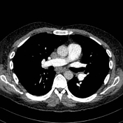
Image 1 CT (selected images) ( create )

Image 2 CT (Iodine Map) ( create )

Image 3 CT (C+ arterial phase) ( create )

Image 4 CT (Monochromatic 50 KeV) ( create )


 Unable to process the form. Check for errors and try again.