
Fetal hydrocephalus
Updates to Article Attributes
Fetal hydrocephalus often refers to an extension of fetal ventriculomegaly where the ventricular dilatation is more severe. It is usually defined when the fetal lateral ventricular diameter is greater than 15 mm 1.
Epidemiology
The estimated incidence is 0.5-3 cases per 1000 live births. There may be a very slight increased female predilection 1011.
Pathology
It can be either obstructive or non-obstructive and each can arise from a number of aetiologies. In a small proportion of cases, it carries a familial X-linked inheritance (congenital X linked-linked hydrocephalus).
Causes
Associations
The vast majority of conditions are associated with other intracranial and cranial anomalies 56. The list includes:
-
central nervous system anomalies: (more common
67 and reported in more than 80% of cases)- aqueductal stenosis: one of the commonest causative associations
- Chiari malformations
- neural tube defect(s)
- Dandy-Walker malformation
- encephalocele
- alobar holoprosencephaly
- posterior fossa cysts
- polymicrogyria
-
non-central nervous system anomalies
- craniofacial
- cleft lip +/- palate
- low set ears
- bilateral optic atrophy
- facial bone anomalies
- acrocephalosyndactylia
- congenital cardiovascular anomalies
- gastrointestinal anomalies
- genitourinary anomalies
- skeletal anomalies
- craniofacial
-
syndromes
-
Meckel-Gruber syndrome
1011 -
Miller-Dieker syndrome
1011
-
Meckel-Gruber syndrome
-
chromosomal anomalies: may be present in ~20% of cases
1011-
trisomy 21
78 -
triploidy
78
-
trisomy 21
See also: congenital syndromes associated with enlarged ventricles.
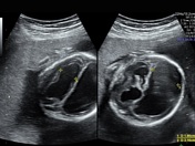
Radiographic features
Antenatal ultrasound
Will demonstrate enlarged ventricles with variable degrees of parenchymal thinning. The choroid may be seen floating within the ventricle giving a dangling choroid sign. Often a separation of more than 3 mm between the choroid plexus and the margin of the ventricle is considered abnormal. In some cases, there may also be evidence of macrocephaly.
Treatment and prognosis
The overall prognosis will depend on the underlying cause and associated anomalies. Some cases can slowly progress during the fetal period. Antenatal shunting has been considered in a small proportion of selected cases 2.
-<p><strong>Fetal hydrocephalus</strong> often refers to an extension of <a href="/articles/fetal-ventriculomegaly">fetal ventriculomegaly</a> where the ventricular dilatation is more severe. It is usually defined when the fetal lateral ventricular diameter is greater than 15 mm <sup>1</sup>.</p><h4>Epidemiology</h4><p>The estimated incidence is 0.5-3 cases per 1000 live births. There may be a very slight increased female predilection <sup>10</sup>.</p><h4>Pathology</h4><p>It can be either obstructive or non-obstructive and each can arise from a number of aetiologies. In a small proportion of cases, it carries a familial X-linked inheritance (<a href="/articles/congenital-x-linked-hydrocephalus">congenital X linked hydrocephalus</a>).</p><h5>Causes</h5><ul><li><a href="/articles/in-utero-infection">in utero infection(s)</a></li></ul><h5>Associations</h5><p>The vast majority of conditions are associated with other intracranial and cranial anomalies <sup>5</sup>. The list includes:</p><ul>- +<p><strong>Fetal hydrocephalus</strong> often refers to an extension of <a href="/articles/fetal-ventriculomegaly">fetal ventriculomegaly</a> where the ventricular dilatation is more severe. It is usually defined when the fetal lateral ventricular diameter is greater than 15 mm <sup>1</sup>.</p><h4>Epidemiology</h4><p>The estimated incidence is 0.5-3 cases per 1000 live births. There may be a very slight increased female predilection <sup>11</sup>.</p><h4>Pathology</h4><p>It can be either obstructive or non-obstructive and each can arise from a number of aetiologies. In a small proportion of cases, it carries a familial X-linked inheritance (<a href="/articles/congenital-x-linked-hydrocephalus">congenital X-linked hydrocephalus</a>).</p><h5>Causes</h5><ul><li><a href="/articles/in-utero-infection">in utero infection(s)</a></li></ul><h5>Associations</h5><p>The vast majority of conditions are associated with other intracranial and cranial anomalies <sup>6</sup>. The list includes:</p><ul>
-<strong>central nervous system anomalies</strong>: (more common <sup>6</sup> and reported in more than 80% of cases)<ul>- +<strong>central nervous system anomalies</strong>: (more common <sup>7</sup> and reported in more than 80% of cases)<ul>
-<a href="/articles/meckel-gruber-syndrome">Meckel-Gruber syndrome</a> <sup>10</sup>- +<a href="/articles/meckel-gruber-syndrome">Meckel-Gruber syndrome</a> <sup>11</sup>
-<a href="/articles/miller-dieker-syndrome">Miller-Dieker syndrome</a> <sup>10</sup>- +<a href="/articles/miller-dieker-syndrome">Miller-Dieker syndrome</a> <sup>11</sup>
-<strong>chromosomal anomalies</strong>: may be present in ~20% of cases <sup>10</sup><ul>- +<strong>chromosomal anomalies</strong>: may be present in ~20% of cases <sup>11</sup><ul>
-<a href="/articles/down-syndrome">trisomy 21</a> <sup>7</sup>- +<a href="/articles/down-syndrome">trisomy 21</a> <sup>8</sup>
-<a href="/articles/triploidy">triploidy</a> <sup>7</sup>- +<a href="/articles/triploidy">triploidy</a> <sup>8</sup>
References changed:
- 5. Cavalheiro S, Moron AF, Zymberg ST et-al. Fetal hydrocephalus--prenatal treatment. Childs Nerv Syst. 2003;19 (7-8): 561-73. <a href="http://dx.doi.org/10.1007/s00381-003-0772-7">doi:10.1007/s00381-003-0772-7</a> - <a href="http://www.ncbi.nlm.nih.gov/pubmed/12908113">Pubmed citation</a><div class="ref_v2"></div>
- 6. Davis GH. Fetal hydrocephalus. Clin Perinatol. 2003;30 (3): 531-9. - <a href="http://www.ncbi.nlm.nih.gov/pubmed/14533894">Pubmed citation</a><div class="ref_v2"></div>
- 7. Pretorius DH, Davis K, Manco-johnson ML et-al. Clinical course of fetal hydrocephalus: 40 cases. AJR Am J Roentgenol. 1985;144 (4): 827-31. <a href="http://www.ajronline.org/cgi/content/abstract/144/4/827">AJR Am J Roentgenol (abstract)</a> - <a href="http://www.ncbi.nlm.nih.gov/pubmed/3883714">Pubmed citation</a><div class="ref_v2"></div>
- 8. Benacerraf BR. Fetal hydrocephalus: diagnosis and significance. Radiology. 1988;169 (3): 858-9. <a href="http://radiology.rsna.org/content/169/3/858.citation">Radiology (citation)</a> - <a href="http://www.ncbi.nlm.nih.gov/pubmed/3055042">Pubmed citation</a><div class="ref_v2"></div>
- 9. Humphreys P, Muzumdar DP, Sly LE et-al. Focal cerebral mantle disruption in fetal hydrocephalus. Pediatr. Neurol. 2007;36 (4): 236-43. <a href="http://dx.doi.org/10.1016/j.pediatrneurol.2006.12.013">doi:10.1016/j.pediatrneurol.2006.12.013</a> - <a href="http://www.ncbi.nlm.nih.gov/pubmed/17437906">Pubmed citation</a><div class="ref_v2"></div>
- 10. Nyberg DA, Mack LA, Hirsch J et-al. Fetal hydrocephalus: sonographic detection and clinical significance of associated anomalies. Radiology. 1987;163 (1): 187-91. <a href="http://radiology.rsna.org/content/163/1/187.abstract">Radiology (abstract)</a> - <a href="http://www.ncbi.nlm.nih.gov/pubmed/3547493">Pubmed citation</a><div class="ref_v2"></div>
- 11. Entezami M, Albig M, Knoll U et-al. Ultrasound Diagnosis of Fetal Anomalies. Thieme. (2003) ISBN:1588902129. <a href="http://books.google.com/books?vid=ISBN1588902129">Read it at Google Books</a> - <a href="http://www.amazon.com/gp/product/1588902129?ie=UTF8&tag=radiopaediaor-20&linkCode=as2&camp=1789&creative=9325&creativeASIN=1588902129">Find it at Amazon</a><div class="ref_v2"></div>
- 4. Cavalheiro S, Moron AF, Zymberg ST et-al. Fetal hydrocephalus--prenatal treatment. Childs Nerv Syst. 2003;19 (7-8): 561-73. <a href="http://dx.doi.org/10.1007/s00381-003-0772-7">doi:10.1007/s00381-003-0772-7</a> - <a href="http://www.ncbi.nlm.nih.gov/pubmed/12908113">Pubmed citation</a><div class="ref_v2"></div>
- 5. Davis GH. Fetal hydrocephalus. Clin Perinatol. 2003;30 (3): 531-9. - <a href="http://www.ncbi.nlm.nih.gov/pubmed/14533894">Pubmed citation</a><div class="ref_v2"></div>
- 6. Pretorius DH, Davis K, Manco-johnson ML et-al. Clinical course of fetal hydrocephalus: 40 cases. AJR Am J Roentgenol. 1985;144 (4): 827-31. <a href="http://www.ajronline.org/cgi/content/abstract/144/4/827">AJR Am J Roentgenol (abstract)</a> - <a href="http://www.ncbi.nlm.nih.gov/pubmed/3883714">Pubmed citation</a><div class="ref_v2"></div>
- 7. Benacerraf BR. Fetal hydrocephalus: diagnosis and significance. Radiology. 1988;169 (3): 858-9. <a href="http://radiology.rsna.org/content/169/3/858.citation">Radiology (citation)</a> - <a href="http://www.ncbi.nlm.nih.gov/pubmed/3055042">Pubmed citation</a><div class="ref_v2"></div>
- 8. Humphreys P, Muzumdar DP, Sly LE et-al. Focal cerebral mantle disruption in fetal hydrocephalus. Pediatr. Neurol. 2007;36 (4): 236-43. <a href="http://dx.doi.org/10.1016/j.pediatrneurol.2006.12.013">doi:10.1016/j.pediatrneurol.2006.12.013</a> - <a href="http://www.ncbi.nlm.nih.gov/pubmed/17437906">Pubmed citation</a><div class="ref_v2"></div>
- 9. Nyberg DA, Mack LA, Hirsch J et-al. Fetal hydrocephalus: sonographic detection and clinical significance of associated anomalies. Radiology. 1987;163 (1): 187-91. <a href="http://radiology.rsna.org/content/163/1/187.abstract">Radiology (abstract)</a> - <a href="http://www.ncbi.nlm.nih.gov/pubmed/3547493">Pubmed citation</a><div class="ref_v2"></div>
- 10. Entezami M, Albig M, Knoll U et-al. Ultrasound Diagnosis of Fetal Anomalies. Thieme. (2003) ISBN:1588902129. <a href="http://books.google.com/books?vid=ISBN1588902129">Read it at Google Books</a> - <a href="http://www.amazon.com/gp/product/1588902129?ie=UTF8&tag=radiopaediaor-20&linkCode=as2&camp=1789&creative=9325&creativeASIN=1588902129">Find it at Amazon</a><div class="ref_v2"></div>
Image 4 Ultrasound (Transverse) ( update )
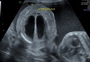
Image 5 Ultrasound ( create )


 Unable to process the form. Check for errors and try again.