
Gastric metastases
Updates to Article Attributes
Gastric metastases are rare, found in less than 2% of patients who die of a carcinoma 6.
Epidemiology
Usually affects the middle-aged and elderly population. Affects males and females equally without predilection.
Clinical presentation
The patient may be asymptomatic, but the most common signs and symptoms include:
- weight loss
- pain
- haematemesis
- melaena
- palpable mass
Pathology
Gastric metastases are usually haematogenous metastases, but the stomach may be involved less frequently by the lymphatic spread or by direct extension of tumour from neighbouring structures or mesenteric reflections.
Primary sites that lead to gastric metastases include the oesophagus, skin (malignant melanoma - sometimes considered the most common 2), lung, cervix, breast, sigmoid colon, testis 1,renal cell carcinoma 7, prostate 8, ovarian adenocarcinoma9, and osteosarcoma (very rare) 10
A solitary metastasis can be much more common than multiple metastases 3.
Location
There may be a predilection for the middle and upper thirds of the stomach 3.
Macroscopic appearance
- may be solitary or multiple
- classic "leather bottle" appearance of the stomach
- polypoid, ulcerated or cavitatory masses
Microscopic appearance
The microscopic features vary greatly depending on the primary cancer.
Radiographic features
Ultrasound
Endoscopic ultrasonography may show a hypoechoic mass disrupting normal wall layers.
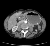
CT
- direct invasion or lymphatic spread to stomach
- distal oesophageal carcinoma
- polypoid, lobulated mass in gastric fundus
- radiologically indistinguishable from primary gastric carcinoma
- pancreatic carcinoma
- pancreatic tumour will be evident
- irregular extrinsic gastric compression
- omental and peritoneal metastases: ovary, uterus, breast, pancreas
- lesions as small as 1 cm can be seen
- lacy reticular pattern to bulky masses /omental cake displace and indent gastric wall
- distal oesophageal carcinoma
- haematogenous spread to stomach
- malignant melanoma
- "target" or bull's-eye lesions, nodular intramural cavitated lesions
- breast cancer: "leather bottle" appearance (linitis plastica)
- markedly thickened gastric wall demonstrating enhancement, preservation of mucosal folds
- mimics primary scirrhous carcinoma of stomach
- malignant melanoma
Treatment and prognosis
Gastric metastases mark advanced disease, and the prognosis is considered poor 1.
Differential diagnosis
- gastritis (erosive type): multiple punctate collections of barium enveloped by thin radiolucent haloes of oedematous mucosa
- pancreatitis
- gastric adenocarcinoma
- gastrointestinal stromal tumour (GIST)
- gastric pseudolymphoma
-</ul><h4>Pathology</h4><p>Gastric metastases are usually haematogenous metastases, but the stomach may be involved less frequently by the lymphatic spread or by direct extension of tumour from neighbouring structures or mesenteric reflections. </p><p>Primary sites that lead to gastric metastases include the oesophagus, skin (<a href="/articles/malignant-melanoma">malignant melanoma</a> - sometimes considered the most common <sup>2</sup>), lung, cervix, breast, sigmoid colon, testis <sup>1</sup>,<sup> </sup><a href="/articles/renal-cell-carcinoma-tnm-staging">renal cell carcinoma </a><sup>7</sup>, prostate <sup>8</sup>, <a href="/articles/ovarian-adenocarcinoma">ovarian adenocarcinoma</a> <sup>9</sup>, and <a href="/articles/osteosarcoma">osteosarcoma</a> (very rare) <sup>10</sup></p><p>A solitary metastasis can be much more common than multiple metastases <sup>3</sup>. </p><h5>Location</h5><p>There may be a predilection for the middle and upper thirds of the <a href="/articles/stomach">stomach</a> <sup>3</sup>.</p><h5>Macroscopic appearance</h5><ul>- +</ul><h4>Pathology</h4><p>Gastric metastases are usually haematogenous metastases, but the stomach may be involved less frequently by the lymphatic spread or by direct extension of tumour from neighbouring structures or mesenteric reflections. </p><p>Primary sites that lead to gastric metastases include the oesophagus, skin (<a href="/articles/malignant-melanoma">malignant melanoma</a> - sometimes considered the most common <sup>2</sup>), lung, cervix, breast, sigmoid colon, testis <sup>1</sup>,<sup> </sup><a href="/articles/renal-cell-carcinoma-tnm-staging">renal cell carcinoma </a><sup>7</sup>, prostate <sup>8</sup>, <a href="/articles/ovarian-adenocarcinoma">ovarian adenocarcinoma</a> <sup>9</sup>, and <a href="/articles/osteosarcoma">osteosarcoma</a> (very rare) <sup>10</sup></p><p>A solitary metastasis can be much more common than multiple metastases <sup>3</sup>. </p><h5>Location</h5><p>There may be a predilection for the middle and upper thirds of the <a href="/articles/stomach">stomach</a> <sup>3</sup>.</p><h5>Macroscopic appearance</h5><ul>
Image 3 CT (C+ portal venous phase) ( update )
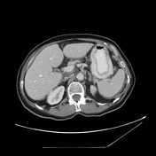
Image 4 CT (C+ arterial phase) ( create )


 Unable to process the form. Check for errors and try again.