
Hepatic hydatid infection
Updates to Article Attributes
Hepatic hydatid disease is a parasitic zoonosis caused by the Echinococcus tapeworm. In the liver, two agents are recognised as causing disease in humans:
- Echinococcus granulosus
- Echinococcus multilocularis
For a general discussion, and for links to other system-specific manifestations, please refer to the article on hydatid disease. For a more specific discussion related to the invasive pattern attributed to the E. multilocularis infection, please refer to the article on alveolar echinococcosis.
Pathology
The parasite E. granulosus is endemic in North America and Australia and is commonly seen in the liver. It typically forms a spherical, fibrous-rimmed cyst with little, if any, surrounding host reaction. Classically it has a large parent cyst within which numerous peripheral daughter cysts are present. Satellite daughter cysts (outside the parent cyst) are seen frequently (~16% cases).There are two forms of E. granulosus:
- pastoral: the most common form; the domestic dog is the main host
- sylvatic: wolf or dog is the main host
The E. multilocularis definitive host (adult parasite) is the red fox (Vulpes vulpes) (sometimes cats and dogs as well), with humans serving as the accidental intermediate host. It is widely distributed throughout the Northern hemisphere.
Radiographic features
This article will discuss the most common presentation of the hepatic hydatid disease, characterised by well-defined encapsulated cystic or multicystic masses related to E. granulosus. For a specific discussion on the less common invasive form, caused by E. multilocularis, please refer to the article on alveolar echinococcosis.
Plain radiograph
May show a curvilinear or ring calcific shadow overlying the liver due to calcification of the pericyst.
Ultrasound
Septated cyst with "daughter" cysts and echogenic material between the cysts. Appearances can vary. May show a double echogenic shadow due to the pericyst. The stage of the cyst may be classified on ultrasound, see: World Health Organization 2001 classification of hepatic hydatid cysts.
CT
Fluid density cyst, with frequent peripheral focal areas of calcification, usually indicates no active infection if completely circumferential. Septa and daughter cysts may be visualised. The water-lily sign indicates a cyst with a floating, undulating membrane, caused by a detached endocyst. May also show hyperdense internal septa within a cyst showing a spoke wheel pattern. Fluid is of variable attenuation, depending on the amount of proteinaceous debris. May show dilated intrahepatic bile ducts due to compression or rupture of the cyst into bile ducts.
MRI
- T1: mixed low signal (depending on the amount of proteinaceous cellular debris)
- T2: mixed high signal (depending on the amount of proteinaceous cellular debris), septa and daughter cysts are well visualised (especially on single-shot T2 sequences)
- T1 C+ (Gd): the walls and septa enhance
Treatment and prognosis
Complications
Rupture into the:
- biliary tree
- peritoneal space (if exophytic)
- bloodstream
- lung 5
See also
-<p><strong>Hepatic hydatid disease</strong> is a parasitic <a title="Zoonosis" href="/articles/zoonosis">zoonosis</a> caused by the <em>Echinococcus </em>tapeworm. In the liver, two agents are recognised as causing disease in humans:</p><ul>- +<p><strong>Hepatic hydatid disease</strong> is a parasitic <a href="/articles/zoonosis">zoonosis</a> caused by the <em>Echinococcus </em>tapeworm. In the liver, two agents are recognised as causing disease in humans:</p><ul>
-</ul>- +</ul><h4><sup>See also</sup></h4><ul><li><a title="Hydatid cyst signs" href="/articles/hydatid-cyst-signs">Hydatid cyst signs</a></li></ul>
Image ( update )
Image ( update )
Image ( update )
Image ( update )
Image ( update )
Image ( update )
Image ( update )
Image ( update )
Image ( update )
Image ( update )
Image ( update )
Image ( update )
Image ( update )
Image 6 Photo ( update )

Image 8 Ultrasound ( update )

Image 9 Ultrasound ( update )

Image 10 CT (non-contrast) ( update )

Image 12 MRI (T2 fat sat) ( update )

Image 14 CT (C+ portal venous phase) ( update )

Image 20 CT (C+ portal venous phase) ( update )

Image 21 MRI (T2) ( update )

Image 22 MRI (T2) ( update )

Image 24 MRI (T2) ( update )

Image 26 CT (C+ portal venous phase) ( update )
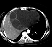
Image 28 CT (non-contrast) ( update )


 Unable to process the form. Check for errors and try again.