
Hyperparathyroidism
Updates to Article Attributes
Body
was changed:
Hyperparathyroidism is the effect of excess parathyroid hormone in the body. It can be primary, secondary, or tertiary. There are many characteristic imaging features, predominantly involving the skeletal system.
Pathology
Increased levels of the parathyroid hormone lead to increased osteoclastic activity. The resultant bone resorption produces cortical thinning (subperiosteal resorption) and osteopenia.
Subtypes
-
primary hyperparathyroidism
-
parathyroid adenoma (~80%)
- multiple parathyroid adenomas (4%) 5
- parathyroid hyperplasia (10-15%) 2,5
- parathyroid carcinoma (1-5%) 4,5
-
parathyroid adenoma (~80%)
-
secondary hyperparathyroidism
- caused by chronic hypocalcaemia with renal osteodystrophy being the most common cause (others include malnutrition, vitamin D deficiency) 4
- results in parathyroid hyperplasia
-
tertiary hyperparathyroidism
- autonomous parathyroid adenoma caused by the chronic overstimulation of hyperplastic glands in renal insufficiency
Associations
Hyperparathyroidism can occur in the context of the following conditions from parathyroid hyperplasia or less commonly multiple parathyroid adenomas 5:
- multiple endocrine neoplasia type I (MEN1)
- multiple endocrine neoplasia type IIa (MEN2a)
- familial hypocalciuric hypercalcaemia
- familial isolated primary hyperparathyroidism
- hyperparathyroidism-jaw tumour syndrome
Radiographic features
-
subperiosteal bone resorption
- classically affects the radial aspects of the proximal and middle phalanges of the 2nd and 3rd fingers
- medial aspect of tibia, femur, humerus
- lamina dura: floating teeth (not specific)
- subchondral resorption
- subligamentous resorption
- ischial tuberosity
- trochanters
- inferior surface of calcaneus and clavicle
- intracortical resorption: cigar/oval-shaped or tunnel-shaped radiolucency in the cortex
- terminal tuft erosion (acro-osteolysis)
- rugger jersey spine
- osteopenia
- brown tumours
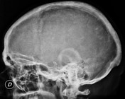
- salt and pepper sign in the skull (pepper pot skull)
- chondrocalcinosis
Findings in secondary (and tertiary) hyperparathyroidism are often associated with the osteosclerosis of renal osteodystrophy and the osteomalacia of vitamin D deficiency:
- subperiosteal bone resorption
- osteopenia
- osteosclerosis, e.g. rugger-jersey spine
- soft tissue calcification
- superscan: generalised increased uptake on Tc-99m pertechnetate bone scan (focal uptake with adenoma)
- superior and inferior rib notching
-<li><a href="/articles/rugger-jersey-spine-hyperparathyroidism">rugger jersey spine</a></li>- +<li><a href="/articles/rugger-jersey-spine-hyperparathyroidism-1">rugger jersey spine</a></li>
Images Changes:
Image 4 X-ray (Lateral) ( create )


 Unable to process the form. Check for errors and try again.