Intra-abdominal calcification
Updates to Article Attributes
Body
was changed:
Intra-abdominal calcification is common and the causes may be classified into four broad groups based on morphology:
Concretions
These are discrete precipitates in a vessel or organ. They are sharp in outline but the density and shape vary but in some cases they may be virtually pathognomonic:
- stones
- pancreatic ductal calcification
- nodal calcification: most commonly from treated lymphoma, tuberculosis or histoplasmosis 1,
- phlebolith
- appendicolith
- calcified granuloma
- failed renal transplant
Conduit calcification
Calcification within the walls of any fluid-filled hollow tube:
- abdominal aorta
- pancreatic ducts
- vas deferens
- large veins
Cystic calcification
Calcification in the wall of a mass such as a cyst, pseudocyst or aneurysm. Hallmark is a smooth curvilinear rim of calcification.
- simple serous cysts
- aneurysms
- echinococcal cysts
- haematoma
- 'porcelain' gallbladder
- calcified appendiceal mucocoele
Solid mass calcification
Diverse features which generally show extensive but variable calcification.
- mesenteric nodes
- adrenal calcifications
- uterine fibroids
- primary tumours, e.g. ovarian dermoid
- metastases
- adenoma
- spleen (autosplenectomy in sickle cell disease)
See also
-<p><a href="/articles/renal-tuberculosis-with-autonephrectomy">Renal tuberculosis with autonephrectomy</a></p>- +<p><a href="/articles/renal-tuberculosis-with-autonephrectomy">renal tuberculosis with autonephrectomy</a></p>
Images Changes:
Image 14 X-ray (Frontal (zoomed in)) ( update )

Caption
was changed:
Case 14: Adrenaladrenal calcification
Image 15 CT (MPR) ( update )
Caption
was changed:
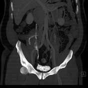
Case 15: Schistosomiasis causing bladderschistosomiasis (bladder and appendix calcification)
Image 16 CT (non-contrast) ( update )

Caption
was changed:
Case 16: Renalrenal tuberculosis with autonephrectomy
Image 17 CT (C+ portal venous phase) ( update )

Caption
was changed:
Case 17: Retroperitonealretroperitoneal hydatid cyst (type III)
Image 18 X-ray (Frontal) ( update )

Caption
was changed:
Case 18: Staghornstaghorn calculus
Image 19 CT (C+ portal venous phase) ( update )

Caption
was changed:
Case 19: Encapsulatingencapsulating peritoneal sclerosis


 Unable to process the form. Check for errors and try again.