
Lung cancer
Updates to Article Attributes
Lung cancer, or frequently, if somewhat incorrectly, known as bronchogenic carcinoma, is the most common cause of cancer in men, and the 6th most frequent cancer in women worldwide. It is the leading cause of cancer mortality worldwide in both men and women and accounts for approximately 20% of all cancer deaths 1.
Epidemiology
Lung cancer is the most common fatal malignancy worldwide both in male and female.
Clinical presentation
Patients with lung cancer may be asymptomatic in up to 50% of cases. Cough and dyspnoea are rather non-specific symptoms that are common amongst those with lung cancer.
Central tumours may result in haemoptysis and peripheral lesions with pleuritic chest pain.
Pneumonia, pleural effusion, wheeze, lymphadenopathy are not uncommon. Other symptoms may be secondary to metastases (brain, liver, bone) or to paraneoplastic syndromes.
Pathology
The term bronchogenic carcinoma is somewhat loosely used to refer to primary malignancies of the lung that are associated with inhaled carcinogens 1 and includes four main histological subtypes. These are broadly divided into non small-cell carcinoma and small cell carcinoma as they are differ clinically in terms of presentation, treatment and prognosis:
-
non small-cell lung cancer (NSCLC) (80%)
-
adenocarcinoma (35%)
- most common cell type overall
- most common in women
- most common cell type in non-smokers but still most patients are smokers
- peripheral
-
squamous cell carcinoma (30%)
- strongly associated with smoking
- most common carcinoma to cavitate
- poor prognosis
-
large-cell carcinoma (15%)
- peripherally located
- very large, usually more than 4 cm
-
adenocarcinoma (35%)
-
small cell carcinoma (20%)
- almost always in smokers
- metastasises early
- most common primary lung malignancy to cause paraneoplastic syndromes and SVC obstruction
- worst prognosis
Other malignant pulmonary neoplasms include lymphoma and sarcoma (rare).
Each subtype has a different radiographic appearance, demographic, and prognosis:
- squamous cell carcinoma of the lung
- adenocarcinoma of the lung
- large cell carcinoma of the lung
- small cell carcinoma of the lung
Risk factors
The major risk factor is cigarette smoking which is implicated in 90% of cases and increase the risk of lung cancer, which can be divided by histological subtype 10:
- squamous cell lung cancer: 11x (men), 15x (women)
- small cell lung cancer: 10x (men), 25x (women)
- large cell lung cancer: 7x (men), 8x (women)
- lung adenocarcinoma: 4x (men and women)
Other risk factors:
- asbestos: 5x increased risk
- occupational exposure: uranium, radon, arsenic, chromium
- diffuse lung fibrosis: 10x increased risk
- chronic obstructive pulmonary disease
Staging
Associations
Various paraneoplastic syndromes can arise in the setting of lung cancer:
- endocrine/metabolic
- SIADH causing hyponatraemia: small-cell sub type
- ACTH secretion (Cushing syndrome): carcinoid and small-cell sub types
- carcinoid syndrome
- gynaecomastia
- adrenal insufficiency (Addison disease): from bilateral metastases 7
- hyperparathyroidism: NSCLC can produce parathyroid hormone (extremely rare) 8
- hypocalcaemia: occurs in the setting of skeletal metastases; especially associated with NSCLC 6
- PTHrp causing hypercalcaemia: squamous cell carcinoma
- neurological
- polyneuropathy
- myelopathy
- limbic encephalitis: particularly associated with SCLC 9
- cerebellar degeneration
- Lambert-Eaton myasthenia syndrome
- other
- finger clubbing
- hypertrophic pulmonary osteoarthropathy (HPOA): squamous cell carcinoma subtype
- nephrotic syndrome
- polymyositis 3
- dermatomyositis 3
- eosinophilia
- acanthosis nigricans
- thrombophlebitis: adenocarcinoma subtype
Treatment and prognosis
Treatment and prognosis varies not only with stage, but also with cell type. In general, surgery, chemotherapy, and radiotherapy are offered according to stage, resectability, operability, and functional status.
Non small cell carcinoma
-
treatment
- operable disease (stage I to IIIA): surgery
- unresectable disease: neoadjuvant chemotherapy, radiotherapy
- advanced disease: palliative combined chemotherapy
-
prognosis (5 year survival rates):
- local (stage I): 55-67%
- locally advanced (stages II-IIIA): 23-40%
- advanced (stages IIIB and IV): 1-3%
Small-cell carcinoma
-
treatment
- limited disease: chemoradiotherapy
- extensive disease: palliative combined chemotherapy
-
prognosis: poor
- limited: 5 year survival rate 15-25%
- extensive: 2 year survival 20% (with palliative combined chemotherapy and supportive care)
See also
-<li><a href="/articles/small-cell-lung-cancer-staging">small cell lung cancer staging</a></li>- +<li><a href="/articles/small-cell-lung-cancer-staging-1">small cell lung cancer staging</a></li>
Image ( destroy )
Image ( update )
Image ( update )
Image ( update )
Image ( update )
Image 3 X-ray (Frontal) ( update )

Image 5 X-ray (Frontal) ( create )

Image 7 CT (C+ portal venous phase) ( update )

Image 8 CT (lung window) ( update )

Image 10 CT (C+ arterial phase) ( update )

Image 11 X-ray (Frontal) ( update )

Image 12 X-ray (Frontal) ( update )

Image 13 CT (C+ arterial phase) ( update )

Image 14 X-ray (Lateral) ( update )

Image 15 CT (lung window) ( update )

Image 16 X-ray ( update )

Image 17 X-ray (Frontal) ( update )

Image 18 CT (lung window) ( update )

Image 19 X-ray (Frontal) ( update )
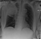
Image 20 X-ray (Frontal) ( update )

Image 21 X-ray (Frontal) ( update )

Image 22 X-ray (Frontal) ( update )

Image 23 CT (renal cortical phase) ( update )

Image 24 CT (C+ delayed) ( update )

Image 25 CT (lung window) ( update )
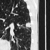
Image 26 CT (lung window) ( update )


 Unable to process the form. Check for errors and try again.