
Senile calcific scleral plaques
Updates to Article Attributes
Senile calcific scleral plaques are benign scleral degenerations common in elderly individuals. They are a common incidental finding on orbital imaging.
Epidemiology
The prevalence of senile scleral calcific plaques increases with age, from ~2-3.5% at age 60, to 25% at age 80 years and over 1,2. They may be more prominent in women than men.
There are no known systemic associations 3.
Clinical presentation
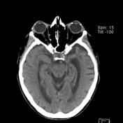
Senile calcific plaques appear as flat, well-circumscribed, ovoid scleral patches. Plaques may appear translucent-blue (due to scleral dehydration and exposure of the underlying uvea), or an opaque grey-white (due to calcification). Approximate dimensions are 2 mm horizontal, 6 mm vertical.
Senile scleral plaques are commonly bilateral and are located in the interpalpebral fissure, 2-3mm posterior to the limbus, and anterior to the insertion of the medial and lateral recti. Plaques involving the vertical muscle insertions are very rare 4,5.
Scleral plaques do not produce symptoms. There are rare reports of extrusion 4.
Pathology
Although the precise aetiology is unknown, calcified senile scleral plaques are considered to represent a form of dystrophic calcification, similar to that which occurs in other parts of the body in areas of hyaline degeneration 6. Various proposed causes include mechanical stress from the rectus muscle insertions, dehydration, and actinic (solar) damage 5.
Histolopathologic evaluation demonstrates calcific deposits within the scleral stroma. These range from fine granular deposits to the confluent plaques which are apparent on imaging. Various staining methods can confirm the presence of calcium, including von Kossa and Alizarin Red stains. Senile calcific plaques were previously thought to represent focal hyalinization of the sclera; however, this has been shown to be false 3,4.
Radiographic features
If sufficiently dense, senile calcific scleral plaques will be visualised on orbital imaging.
Computed Tomography
CT
Scleral plaques appear as small ovoid calcifications along the anterior globe, typically anterior to the horizontal insertion of the rectus muscles 1-3.
Ultrasound
May show anterior shadowing typical of calcified lesions 6.
Optical Coherence Tomography
coherence tomographySenile scleral plaques are hyporeflective on OCT, with hyperreflective calcifications.
Treatment and prognosis
No treatment is required.
Differential diagnosis
Although scleral calcification may occur in the setting of inflammation, lymphoma, and hypercalcemic states, senile scleral calcifications are generally considered "do not touch" lesions given their commonality stereotypical appearance.
A number of other conditions cause calcification of the globe.:
- foreign body
- atypical shape or location of classification
- should recommend clinical correlation with physical/ophthalmologic evaluation
-
scleral buckle
- distinct appearance, often encircling, are located more posteriorly, and are placed between the muscle and the globe
- scleromalacia perforans (rare)
- progressive thinning occurring in younger patients
- other pigmented ocular surface lesions
See also
-<p><strong>Senile calcific scleral plaques </strong>are benign scleral degenerations common in elderly individuals. They are a common incidental finding on orbital imaging.</p><h4>Epidemiology</h4><p>The prevalence of senile scleral calcific plaques increases with age, from ~2-3% at age 60, to 25% at age 80 years and over<sup> 1,2</sup>. They may be more prominent in women than men.</p><p>There are no known systemic associations<sup> 3</sup>.</p><h4>Clinical presentation</h4><p>Senile calcific plaques appear as flat, well-circumscribed, ovoid scleral patches. Plaques may appear translucent-blue (due to scleral dehydration and exposure of the underlying uvea), or an opaque grey-white (due to calcification). Approximate dimensions are 2 mm horizontal, 6 mm vertical.</p><p>Senile scleral plaques are commonly bilateral and are located in the interpalpebral fissure, 2-3mm posterior to the limbus, and anterior to the insertion of the medial and lateral recti. Plaques involving the vertical muscle insertions are very rare <sup>4,5</sup>.</p><p>Scleral plaques do not produce symptoms. There are rare reports of extrusion<sup> 4</sup>.</p><h4>Pathology</h4><p>Although the precise aetiology is unknown, calcified senile scleral plaques are considered to represent a form of dystrophic calcification, similar to that which occurs in other parts of the body in areas of hyaline degeneration <sup>6</sup>. Various proposed causes include mechanical stress from the rectus muscle insertions, dehydration, and actinic (solar) damage <sup>5</sup>.</p><p>Histolopathologic evaluation demonstrates calcific deposits within the scleral stroma. These range from fine granular deposits to the confluent plaques which are apparent on imaging. Various staining methods can confirm the presence of calcium, including von Kossa and Alizarin Red stains. Senile calcific plaques were previously thought to represent focal hyalinization of the sclera; however, this has been shown to be false<sup> 3,4</sup>.</p><p> </p><h4>Radiographic features</h4><p>If sufficiently dense, senile calcific scleral plaques will be visualised on orbital imaging.</p><h6>Computed Tomography</h6><p>Scleral plaques appear as small ovoid calcifications along the anterior globe, typically anterior to the horizontal insertion of the rectus muscles<sup> 1-3</sup>.</p><h6>Ultrasound</h6><p>May show anterior shadowing typical of calcified lesions<sup> 6.</sup></p><h6>Optical Coherence Tomography</h6><p>Senile scleral plaques are hyporeflective on OCT, with hyperreflective calcifications.</p><h4>Treatment and prognosis</h4><p>No treatment is required.</p><h4>Differential diagnosis</h4><p>Although scleral calcification may occur in the setting of inflammation, lymphoma, and hypercalcemic states, senile scleral calcifications are generally considered "do not touch" lesions given their commonality stereotypical appearance.</p><p>A number of other conditions cause <a href="/articles/calcification-of-the-globe-differential">calcification of the globe</a>.</p><ul>- +<p><strong>Senile calcific scleral plaques </strong>are benign scleral degenerations common in elderly individuals. They are a common incidental finding on orbital imaging.</p><h4>Epidemiology</h4><p>The prevalence of senile scleral calcific plaques increases with age, from ~2.5% at age 60, to 25% at age 80 years and over<sup> 1,2</sup>. They may be more prominent in women than men.</p><p>There are no known systemic associations<sup> 3</sup>.</p><h4>Clinical presentation</h4><p>Senile calcific plaques appear as flat, well-circumscribed, ovoid scleral patches. Plaques may appear translucent-blue (due to scleral dehydration and exposure of the underlying uvea), or an opaque grey-white (due to calcification). Approximate dimensions are 2 mm horizontal, 6 mm vertical.</p><p>Senile scleral plaques are commonly bilateral and are located in the interpalpebral fissure, 2-3mm posterior to the limbus, and anterior to the insertion of the medial and lateral recti. Plaques involving the vertical muscle insertions are very rare <sup>4,5</sup>.</p><p>Scleral plaques do not produce symptoms. There are rare reports of extrusion<sup> 4</sup>.</p><h4>Pathology</h4><p>Although the precise aetiology is unknown, calcified senile scleral plaques are considered to represent a form of dystrophic calcification, similar to that which occurs in other parts of the body in areas of hyaline degeneration <sup>6</sup>. Various proposed causes include mechanical stress from the rectus muscle insertions, dehydration, and actinic (solar) damage <sup>5</sup>.</p><p>Histolopathologic evaluation demonstrates calcific deposits within the scleral stroma. These range from fine granular deposits to the confluent plaques which are apparent on imaging. Various staining methods can confirm the presence of calcium, including von Kossa and Alizarin Red stains. Senile calcific plaques were previously thought to represent focal hyalinization of the sclera; however, this has been shown to be false<sup> 3,4</sup>.</p><h4>Radiographic features</h4><p>If sufficiently dense, senile calcific scleral plaques will be visualised on orbital imaging.</p><h5>CT</h5><p>Scleral plaques appear as small ovoid calcifications along the anterior globe, typically anterior to the horizontal insertion of the rectus muscles<sup> 1-3</sup>.</p><h5>Ultrasound</h5><p>May show anterior shadowing typical of calcified lesions<sup> 6.</sup></p><h5>Optical coherence tomography</h5><p>Senile scleral plaques are hyporeflective on OCT, with hyperreflective calcifications.</p><h4>Treatment and prognosis</h4><p>No treatment is required.</p><h4>Differential diagnosis</h4><p>Although scleral calcification may occur in the setting of inflammation, lymphoma, and hypercalcemic states, senile scleral calcifications are generally considered "do not touch" lesions given their commonality stereotypical appearance.</p><p>A number of other conditions cause <a href="/articles/calcification-of-the-globe-differential">calcification of the globe</a>:</p><ul>
-</ul><p> </p><h4>See also</h4><p><a href="/articles/calcification-of-the-globe-differential">Calcification of the globe (differential)</a></p>- +</ul><h4>See also</h4><p><a href="/articles/calcification-of-the-globe-differential">Calcification of the globe (differential)</a></p>
Tags changed:
- eye
- globe
Image 1 CT (non-contrast) ( update )


 Unable to process the form. Check for errors and try again.