
Subiculum
Updates to Article Attributes
The subiculum is located in the mesial temporal lobe and is a subdivision of the hippocampal formation, along with Ammon’s horn, the entorhrinal cortex and the hippocampus proper. It is the predominant output source of the hippocampal formation.
Structure
The subiculum occupies a portion of the parahippocampal gyrus in the mesial temporal lobe and is a component of the medial temporal memory system. It is made up of several cortical fields and for this reason is sometimes referred to as the subicular complex. It is positioned between the CA1 region of the hippocampus proper and the entorhinal cortex ventrally. Dorsally, it is bordered by the retrosplenial cortex.
The subicular complex is broken down into several distinct but related areas by their differing cytoarchitecture, however with slight variations between human, monkey and rodent studies. These areas include the prosubiculum (ProS), subiculum proper (Sub), presubiculum (PrS), postsubiculum (PoS), and parasubiculum (PaS)
Sub: The subiculum proper is bordered proximally by the ProS, which separates it from the CA1 region of the hippocampus. Distally, it is bordered by the PreS.
ProS: The area separating the subiculum proper from the CA1 region of the hippocampus proper.
PrS: This area borders the subiculum proper ventrally and makes up part of the parahippocampal gyrus.
PoS: The PoS is bordered by the presubiculum ventrally and laterally, with the border between the two characterised by an abrupt change in cyto and histochemical staining. However, some sources treat this area as the dorsal part of the presubiculum.
PaS: This area forms the ventral shoulder of the parahippocampal gyrus and continues the PrS ventrally. It is bordered laterally by the entorhinal cortex.
Radiographic Featuresfeatures
MRI
As with the hippocampus proper, the subiculum is best visualised on coronal plane MR images. A detailed description of associated landmarks is available in the hippocampus article.
Clinical Significancesignificance
There is ongoing emerging evidence of the role of the subiculum in Alzheimer’s disease and epilepsy.
Etymology:History and etymology
The name subiculum is derived from the Latin word for support. This reflects the location of the subiculum under the hippocampus proper.
-</xml><![endif]--></p><p>The <strong>subiculum</strong> is located in the <a title="Mesial temporal lobe" href="/articles/mesial-temporal-lobe">mesial temporal lobe</a> and is a subdivision of the <a title="Hippocampus" href="/articles/hippocampus">hippocampal formation</a>, along with <a title="Ammon’s horn" href="/articles/ammon-s-horn">Ammon’s horn</a>, the <a title="Entorhinal cortex" href="/articles/entorhinal-cortex">entorhrinal cortex</a> and the <a title="Hippocampus" href="/articles/hippocampus">hippocampus</a> proper. It is the predominant output source of the hippocampal formation.</p><h4>Structure</h4><p>The subiculum occupies a portion of the <a title="Parahippocampal gyrus" href="/articles/parahippocampal-gyrus">parahippocampal gyrus</a> in the <a title="Mesial temporal lobe" href="/articles/mesial-temporal-lobe">mesial temporal lobe</a> and is a component of the medial temporal memory system. It is made up of several cortical fields and for this reason is sometimes referred to as the subicular complex. It is positioned between the CA1 region of the <a title="Hippocampus" href="/articles/hippocampus">hippocampus</a> proper and the <a title="Entorhinal cortex" href="/articles/entorhinal-cortex">entorhinal cortex</a> ventrally. Dorsally, it is bordered by the <a title="retrosplenial cortex" href="/articles/retrosplenial-cortex">retrosplenial cortex</a>.</p><p>The subicular complex is broken down into several distinct but related areas by their differing cytoarchitecture, however with slight variations between human, monkey and rodent studies. These areas include the prosubiculum (ProS), subiculum proper (Sub), presubiculum (PrS), postsubiculum (PoS), and parasubiculum (PaS)</p><ul>-<li>Sub: The subiculum proper is bordered proximally by the ProS, which separates it from the CA1 region of the <a title="Hippocampus" href="/articles/hippocampus">hippocampus</a>. Distally, it is bordered by the PreS.</li>-<li>ProS: The area separating the subiculum proper from the CA1 region of the hippocampus proper.</li>-<li>PrS: This area borders the subiculum proper ventrally and makes up part of the <a title="Parahippocampal gyrus" href="/articles/parahippocampal-gyrus">parahippocampal gyrus</a>.</li>-<li>PoS: The PoS is bordered by the presubiculum ventrally and laterally, with the border between the two characterised by an abrupt change in cyto and histochemical staining. However, some sources treat this area as the dorsal part of the presubiculum.</li>-<li>PaS: This area forms the ventral shoulder of the parahippocampal gyrus and continues the PrS ventrally. It is bordered laterally by the <a title="Entorhinal cortex" href="/articles/entorhinal-cortex">entorhinal cortex.</a>-</li>-</ul><h4>Radiographic Features</h4><h4>MRI</h4><p>As with the <a title="Hippocampus" href="/articles/hippocampus">hippocampus</a> proper, the subiculum is best visualised on coronal plane MR images. A detailed description of associated landmarks is available in the <a title="Hippocampus" href="/articles/hippocampus">hippocampus</a> article.</p><h4>Clinical Significance</h4><p>There is ongoing emerging evidence of the role of the subiculum in <a title="Alzheimer disease" href="/articles/alzheimer-disease-1">Alzheimer’s</a> disease and <a title="Epilepsy" href="/articles/epilepsy">epilepsy</a>.</p><h4>Etymology:</h4><p>The name subiculum is derived from the Latin word for support. This reflects the location of the subiculum under the <a title="Hippocampus" href="/articles/hippocampus">hippocampus</a> proper.</p><p><!--[if gte mso 9]><xml>- +</xml><![endif]--></p><p>The <strong>subiculum</strong> is located in the <a href="/articles/mesial-temporal-lobe">mesial temporal lobe</a> and is a subdivision of the <a href="/articles/hippocampus">hippocampal formation</a>, along with <a href="/articles/ammon-s-horn">Ammon’s horn</a>, the <a href="/articles/entorhinal-cortex">entorhrinal cortex</a> and the <a href="/articles/hippocampus">hippocampus</a> proper. It is the predominant output source of the hippocampal formation.</p><h4>Structure</h4><p>The subiculum occupies a portion of the <a href="/articles/parahippocampal-gyrus">parahippocampal gyrus</a> in the <a href="/articles/mesial-temporal-lobe">mesial temporal lobe</a> and is a component of the medial temporal memory system. It is made up of several cortical fields and for this reason is sometimes referred to as the subicular complex. It is positioned between the CA1 region of the <a href="/articles/hippocampus">hippocampus</a> proper and the <a href="/articles/entorhinal-cortex">entorhinal cortex</a> ventrally. Dorsally, it is bordered by the <a href="/articles/retrosplenial-cortex">retrosplenial cortex</a>.</p><p>The subicular complex is broken down into several distinct but related areas by their differing cytoarchitecture, however with slight variations between human, monkey and rodent studies. These areas include the prosubiculum (ProS), subiculum proper (Sub), presubiculum (PrS), postsubiculum (PoS), and parasubiculum (PaS)</p><p><strong>Sub</strong>: The subiculum proper is bordered proximally by the ProS, which separates it from the CA1 region of the <a href="/articles/hippocampus">hippocampus</a>. Distally, it is bordered by the PreS.</p><p><strong>ProS</strong>: The area separating the subiculum proper from the CA1 region of the hippocampus proper.</p><p><strong>PrS</strong>: This area borders the subiculum proper ventrally and makes up part of the <a href="/articles/parahippocampal-gyrus">parahippocampal gyrus</a>.</p><p><strong>PoS</strong>: The PoS is bordered by the presubiculum ventrally and laterally, with the border between the two characterised by an abrupt change in cyto and histochemical staining. However, some sources treat this area as the dorsal part of the presubiculum.</p><p><strong>PaS</strong>: This area forms the ventral shoulder of the parahippocampal gyrus and continues the PrS ventrally. It is bordered laterally by the <a href="/articles/entorhinal-cortex">entorhinal cortex.</a></p><h4>Radiographic features</h4><h4>MRI</h4><p>As with the <a href="/articles/hippocampus">hippocampus</a> proper, the subiculum is best visualised on coronal plane MR images. A detailed description of associated landmarks is available in the <a href="/articles/hippocampus">hippocampus</a> article.</p><h4>Clinical significance</h4><p>There is ongoing emerging evidence of the role of the subiculum in <a href="/articles/alzheimer-disease-1">Alzheimer’s</a> disease and <a href="/articles/epilepsy">epilepsy</a>.</p><h4>History and etymology</h4><p>The name subiculum is derived from the Latin word for support. This reflects the location of the subiculum under the <a href="/articles/hippocampus">hippocampus</a> proper.</p><p><!--[if gte mso 9]><xml>
References changed:
- 1. Susan Standring. Gray's Anatomy. ISBN: 9780702052309
- 2. O'Mara S. The subiculum: what it does, what it might do, and what neuroanatomy has yet to tell us. Journal of anatomy. 207 (3): 271-82. <a href="https://doi.org/10.1111/j.1469-7580.2005.00446.x">doi:10.1111/j.1469-7580.2005.00446.x</a> - <a href="https://www.ncbi.nlm.nih.gov/pubmed/16185252">Pubmed</a> <span class="ref_v4"></span>
- 3. Insausti R, Muñoz-López M, Insausti AM, Artacho-Pérula E. The Human Periallocortex: Layer Pattern in Presubiculum, Parasubiculum and Entorhinal Cortex. A Review. Frontiers in neuroanatomy. 11: 84. <a href="https://doi.org/10.3389/fnana.2017.00084">doi:10.3389/fnana.2017.00084</a> - <a href="https://www.ncbi.nlm.nih.gov/pubmed/29046628">Pubmed</a> <span class="ref_v4"></span>
- 4. Aggleton JP, Christiansen K. The subiculum: the heart of the extended hippocampal system. Progress in brain research. 219: 65-82. <a href="https://doi.org/10.1016/bs.pbr.2015.03.003">doi:10.1016/bs.pbr.2015.03.003</a> - <a href="https://www.ncbi.nlm.nih.gov/pubmed/26072234">Pubmed</a> <span class="ref_v4"></span>
- Susan Standring. Gray's Anatomy. ISBN: 9780702052309
- O'Mara S. The subiculum: what it does, what it might do, and what neuroanatomy has yet to tell us. Journal of anatomy. 207 (3): 271-82. <a href="https://doi.org/10.1111/j.1469-7580.2005.00446.x">doi:10.1111/j.1469-7580.2005.00446.x</a> - <a href="https://www.ncbi.nlm.nih.gov/pubmed/16185252">Pubmed</a> <span class="ref_v4"></span>
- Insausti R, Muñoz-López M, Insausti AM, Artacho-Pérula E. The Human Periallocortex: Layer Pattern in Presubiculum, Parasubiculum and Entorhinal Cortex. A Review. Frontiers in neuroanatomy. 11: 84. <a href="https://doi.org/10.3389/fnana.2017.00084">doi:10.3389/fnana.2017.00084</a> - <a href="https://www.ncbi.nlm.nih.gov/pubmed/29046628">Pubmed</a> <span class="ref_v4"></span>
- Aggleton JP, Christiansen K. The subiculum: the heart of the extended hippocampal system. Progress in brain research. 219: 65-82. <a href="https://doi.org/10.1016/bs.pbr.2015.03.003">doi:10.1016/bs.pbr.2015.03.003</a> - <a href="https://www.ncbi.nlm.nih.gov/pubmed/26072234">Pubmed</a> <span class="ref_v4"></span>
Image 1 Diagram ( update )
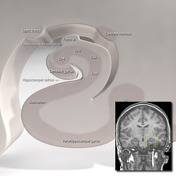
Image 2 MRI (T2) ( update )


 Unable to process the form. Check for errors and try again.