
Temporal bone
Updates to Article Attributes
Body
was changed:
The temporal bone is situated on the sides and the base of the cranium and lateral to the temporal lobe of the cerebrum. The temporal bone is one of the most important calvarial and skull base bones.
Gross anatomy
The temporal bone is very complex and consists of five parts 1,2:
Some anatomists describe only 4 parts of the temporal bone, with the mastoid and petrous parts combined as a petromastoid part 3.
The temporal bone can also be divided into otologic zones:
- the medial third of the external auditory canal (part of the external ear)
- middle ear (tympanic cavity)
- inner ear
- internal auditory canal
Articulations
There are four intrinsic fissures between the divisions of the temporal bone 4,5:
There are many named extrinsic fissures between the temporal bone and other cranial bones 4,5:
-<li>the medial third of the <a title="External auditory canal" href="/articles/external-auditory-canal">external auditory canal</a> (part of the <a href="/articles/external-ear">external ear</a>)</li>-<li><a title="Middle ear" href="/articles/middle-ear">middle ear (tympanic cavity)</a></li>-<li><a title="Inner ear" href="/articles/inner-ear">inner ear</a></li>-<li><a title="Internal auditory canal (IAC)" href="/articles/internal-acoustic-canal">internal auditory canal</a></li>- +<li>the medial third of the <a href="/articles/external-auditory-canal">external auditory canal</a> (part of the <a href="/articles/external-ear">external ear</a>)</li>
- +<li><a href="/articles/middle-ear">middle ear (tympanic cavity)</a></li>
- +<li><a href="/articles/inner-ear">inner ear</a></li>
- +<li><a href="/articles/internal-acoustic-canal">internal auditory canal</a></li>
Images Changes:
Image ( update )
Caption
was changed:
Figure 37: diagram - medial temporal bone
Position
was set to
.
Image 4 CT (Axial Annotated) ( create )

Caption
was changed:
Figure 2: temporal bone structures (CT anatomy(axial)
Image 5 CT (Coronal Annotated) ( create )

Image 6 CT (Stenvers Annotated) ( create )
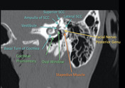
Image 7 CT (Labelled) ( update )

Caption
was changed:
Figure 45: normal petrous temporal bone axial CT
Position
was set to
.
Image 8 Diagram ( update )

Caption
was changed:
Figure 26: diagram - skull and facial bones
Position
was set to
.


 Unable to process the form. Check for errors and try again.