
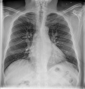
Upper lobe pulmonary venous diversion
Updates to Article Attributes
Upper lobe pulmonary venous diversion (cephalisation(also described as cephalisation of the pulmonary veins) reflects elevation of left atrial pressure and can occur withis an early sign of pulmonary oedema. It produces stag-antler's sign on a frontal chest x-ray.
Clinical presentation
The normal left atrial pressure is 5-10 mmHg. An elevation of left atrial pressure to 10-15 mmHg will result in cephalisationupper lobe venous diversion. Possible causes Typically, the cause for cephalisation include 2:
-
that increase in left atrial pressure is left heart failure (most
commoncommonly) - , or mitral valve disease
shunt
Radiographic features
The comparison vessels shouldOn frontal chest radiograph, it is seen as dilation of the upper lobe pulmonary veins as they branch superiorly from the hilum. This can be measured objectively, with cephalisation present if the upper lobe veins are of the same or higher diameter than the lower lobe veins, measured equidistant from the hilar point2.
See also
-<p><strong>Upper lobe pulmonary venous diversion (cephalisation)</strong> reflects elevation of left atrial pressure and can occur with <a href="/articles/pulmonary-oedema">pulmonary oedema</a>. It produces <a href="/articles/stags-antler-sign-lungs">stag-antler's sign</a> on a frontal chest x-ray.</p><p>The normal left atrial pressure is 5-10 mmHg. An elevation of left atrial pressure to 10-15 mmHg will result in cephalisation. Possible causes for cephalisation include <sup>2</sup>:</p><ul>-<li>-<a href="/articles/left-heart-failure">left heart failure</a> (most common)</li>-<li><a href="/articles/mitral-valve-disease">mitral valve disease</a></li>-<li>shunt</li>-</ul><p>The comparison vessels should be equidistant from the <a href="/articles/hilar-point">hilar point</a>. </p>- +<p><strong>Upper lobe pulmonary venous diversion </strong>(also described as <strong>cephalisation of the pulmonary veins</strong>) reflects elevation of left atrial pressure and is an early sign of <a href="/articles/pulmonary-oedema">pulmonary oedema</a>.</p><h4>Clinical presentation</h4><p>The normal left atrial pressure is 5-10 mmHg. An elevation of left atrial pressure to 10-15 mmHg will result in upper lobe venous diversion. Typically, the cause for that increase in left atrial pressure is <a href="/articles/left-heart-failure">left heart failure</a> (most commonly), or <a title="Mitral valve disease" href="/articles/mitral-valve-disease">mitral valve disease</a><span> <sup>1</sup>.</span></p><h4>Radiographic features</h4><p>On frontal chest radiograph, it is seen as dilation of the upper lobe <a title="Pulmonary veins" href="/articles/pulmonary-veins">pulmonary veins</a> as they branch superiorly from the hilum. This can be measured objectively, with cephalisation present if the upper lobe veins are of the same or higher diameter than the lower lobe veins, measured equidistant from the <a title="Hilar point" href="/articles/hilar-point">hilar point</a> <sup>2</sup>.</p><h4>See also</h4><ul><li><a title="Stag's antler sign (lungs)" href="/articles/stags-antler-sign-lungs">stag's antler sign</a></li></ul>
References changed:
- 1. Goodman LR, Felson B. Felson's Principles of Chest Roentgenology. W.B. Saunders Company. (2007) ISBN:1416029230. <a href="http://books.google.com/books?vid=ISBN1416029230">Read it at Google Books</a> - <a href="http://www.amazon.com/gp/product/1416029230?ie=UTF8&tag=radiopaediaor-20&linkCode=as2&camp=1789&creative=9325&creativeASIN=1416029230">Find it at Amazon</a><div class="ref_v2"></div>
- 2. Barile M. Pulmonary Edema: A Pictorial Review of Imaging Manifestations and Current Understanding of Mechanisms of Disease. (2020) European journal of radiology open. 7: 100274. <a href="https://doi.org/10.1016/j.ejro.2020.100274">doi:10.1016/j.ejro.2020.100274</a> - <a href="https://www.ncbi.nlm.nih.gov/pubmed/33163585">Pubmed</a> <span class="ref_v4"></span>
- 4. Lacey GD, Morley S, Berman L. The Chest X-Ray, A Survival Guide. Saunders. (2008) ISBN:0702030465. <a href="http://books.google.com/books?vid=ISBN0702030465">Read it at Google Books</a> - <a href="http://www.amazon.com/gp/product/0702030465?ie=UTF8&tag=radiopaediaor-20&linkCode=as2&camp=1789&creative=9325&creativeASIN=0702030465">Find it at Amazon</a><div class="ref_v2"></div>
- 1. Lacey GD, Morley S, Berman L. The Chest X-Ray, A Survival Guide. Saunders. (2008) ISBN:0702030465. <a href="http://books.google.com/books?vid=ISBN0702030465">Read it at Google Books</a> - <a href="http://www.amazon.com/gp/product/0702030465?ie=UTF8&tag=radiopaediaor-20&linkCode=as2&camp=1789&creative=9325&creativeASIN=0702030465">Find it at Amazon</a><div class="ref_v2"></div>
- 2. Goodman LR, Felson B. Felson's Principles of Chest Roentgenology. W.B. Saunders Company. (2007) ISBN:1416029230. <a href="http://books.google.com/books?vid=ISBN1416029230">Read it at Google Books</a> - <a href="http://www.amazon.com/gp/product/1416029230?ie=UTF8&tag=radiopaediaor-20&linkCode=as2&camp=1789&creative=9325&creativeASIN=1416029230">Find it at Amazon</a><div class="ref_v2"></div>
Image 3 X-ray (Frontal) ( create )


 Unable to process the form. Check for errors and try again.