Radiopaedia Blog

Today we're announcing a "bonus video" offer. Register before March 31 for our Emergency Radiology Course and you'll receive bonus access to a 30-minute pediatric emergency radiology video recorded by Dr Jeremy Jones (UK).

This bonus offer applies to anyone who registers to attend the course in Melbourne on May 16 and also to anyone who registers to watch the course online. The cost to watch online is just $25 which is amazingly good value, and for people in 116 developing countries the course is free! 

If you've already registered for the Emergency Radiology Course then you don't need to do anything; we'll automatically give you access to the "bonus video" as soon as it is available.

Key dates
  • March 31 - last day to qualify for the "bonus video"
  • Early April - "bonus video" available for viewing
  • May 16 - full course takes place in Melbourne
  • May 30 - last day to register to watch course online
  • June 1 - full course video available for viewing


10th Mar 2015 11:03 UTC

Goodbye and thank you Ray

It is with great sadness that I found out today, rather belatedly, that one of our past editors, J. Ray Ballinger, passed last year. Ray was one of the very first to embrace online education, starting MRItutor.org in 1994, and as such is one of the fathers of what we now recognize as open-access education. 

In 2007, shortly after opening Radiopaedia.org to the world I reached out to Ray and asked if he would be interested in contributing. He replied:

Hi Frank,
Let me give that some thought.
Regards,
Ray

True to his word he did. Six years later, in 2013, I unexpectedly received an email from Ray. 

Hi Frank,

I was looking at your site again and have decided, rather belatedly, to offer any of my mritutor.org website materials to yours. I retired last year a bit early than I anticipated because of treatment for stage IV carcinoid cancer and may not be around in a few years to continue it. I can fill in some of the physics topics that you need as well as add or modify some of your existing articles if you wish. [...]

Warm wishes,

Ray

Needless to say, I was terribly moved. That someone would care so much about teaching, at what must have been such a difficult time, was very inspiring. Ray became our physics editor during which time he made over 170 edits, many on improving the large contribution imported into Radiopaedia.org from his site. 

During this time we exchanged many emails. I found Ray to be a funny and candid person, who clearly cared deeply about the idea of creating educational material and sharing it with the world. 

We will all miss Ray, and can all learn a great deal from the example he has set. 


Frank Gaillard

Founder and Editor, Radiopaedia.org

Radiopaedia.org is fundamentally about facilitating access to quality radiology teaching material, so that medical professionals from around the world can better diagnose and treat their patients. I believe it is crucial that such access is not determined by the wealth of your country or that of your institution. 

Unfortunately much of the medical education industry (journals, textbooks, courses and meetings) functions primarily by directly monetizing access to this information. Although we have made great inroads in creating a free resource of thousands of articles and cases, we recognize the importance of meetings where an expert can guide you through the diagnostic process. This is one of the reasons why we have started running Radiopaedia Courses and making these course as easily available as possible. We can't do it all on our own however. 

Thus, when I was approached by the organizing committee of ANZSNR 2015 (Australian and New Zealand Society of Neuroradiology - March 21-22 - Melbourne, Australia) to help bring at least some of the meeting  to the world, I was wrapped. Together with my colleagues A.Prof Peter Mitchell, A.Prof Pramit Phal and Dr Christine Goh, we will be presenting over 5 hours of case-based learning, covering the spine, cerebrovascular conditions, pitutiary region, tumors and tumor like conditions, and neurodegenerative diseases. 

I have had the pleasure of learning a great deal from all my fellow speakers over the years, and can say without equivocation that watching them explain their approach to a case and how they narrow down a differential is an exceptional way to learn.  

It is therefore with great pleasure that we can share with you over 100 of the cases that we will be presenting and discussing at the meeting. Radiology is not about the answer, so much as the process, so feel free to read through these cases ahead of the meeting.

For those of you not able to attend, I hope you will gain a great deal from this collection of curated cases, and that you won't feel too bad about missing out on the live event; there is always next year.  :)

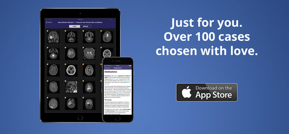
In addition to accessing these cases through your browser (on any computer or device), we have also created a bespoke iOS (iPhone / iPad). 

To find the app (free) just follow this link, or merely search for "ANZSNR" in the App Store. 

To view the cases within Radiopaedia.org, follow (and bookmark) the links below. 

If you are lucky enough to be able to attend ANZSNR 2015 (meeting details here), and you see me milling about, please come up and say hi and let me know what you think of the app / cases. 

Frank Gaillard

Dr Frank Gaillard is a neuroradiologist at the Royal Melbourne Hospital, Melbourne, Australia, and is the Founder and Editor of Radiopaedia.org. 

NB: Opinions expressed are those of the author alone, and are not those of his employer, or of Radiopaedia.org


To whet your appetite for our upcoming Emergency Radiology Course - which you can watch online for just $25 - I thought I'd share three classic conditions that anyone working in the ER should be able to diagnose on CT. 

Appendicitis

The left hand coronal CT image shows a blind ending tubular structure in the right iliac fossa with a calcified rounded density at its base. This a fairly classic appearance of appendicitis with an appendicolith. While the appendix is mildly distended in this case, there is only minimal inflammatory stranding and no suggestion of perforation or abscess formation. You can read more and see other case examples here.

Pulmonary Embolism

The center axial image from a CT pulmonary angiogram shows a rounded filling defect occluding the left lower lobe pulmonary artery origin. Learning how to track the pulmonary arteries on a CTPA and detect thrombus like this one is a very handy skill. Our pulmonary embolism article has plenty of cases that you can practice on.

Hemorrhagic Stroke - Hypertensive

The right hand axial image of the brain shows a sizable hyperdense region within the left basal ganglia (the lentiform nucleus) consistent with acute intracerebral hemorrhage. This is a classic location for a hemorrhagic stroke secondary to hypertension. This should not be confused with hemorrhagic trasnformation of an ischemic stroke which is a separate entity. Below is Frank Gaillard's Radiology Channel video which covers this topic really well. 


A little while ago I wrote about some of the reasons I thought it was important to measure actual ADC values, rather than just claiming there 'was' or 'wasn't' restricted diffusion. That article used epidermoid cysts to point out why such dichotomous terminology was unhelpful and in some situations confusing.  If you haven't read that post, then I suggest doing so now before we go on. 

Reporting astrocytoma MRI scans is another really common situation in which talking about the 'presence' or 'absence' of restricted diffusion does not makes much sense. It is made even worse by the fact that trying to work if there is significant diffusion restriction (whatever that may mean) is visually very difficult. 

Why is saying there 'is' or 'isn't' restricted diffusion silly? Well, we know that lower ADC values correlated fairly well with histological tumor grade; the higher cellularity, smaller extracellular space seen in high grade tumors leads to greater diffusion restriction. In diffuse astrocytomas, the following values have been reported 1

  • Gd II: 1273 ± 293 10-6 mm2/s
  • Gd III: 1067 ± 276 10-6 mm2/s
  • Gd IV: 745 ± 135 10-6 mm2/s

Figure 1. Astrocytoma (GBM) involving the splenium, has a solid component with lower ADC values than the surrounding white matter, but also has a component of T2 shine-through. Working out how much restricted diffusion is present in the solid component is hard.  Complete case here


So, despite some overlap, this is really quite useful right? Well it is if you bother to measure the values. What isn't useful is looking at the DWI and ADC maps and trying to eyeball the presence or absence of restricted diffusion. For some time I have tried to estimate ADC values of the solid component of a tumor by visually comparing it to normal white matter; I figured that if I could do this reliably then this would be good enough. Unfortunately, there are a number of challenges in accomplishing this visual task. Firstly on DWI there is invariably a component of T2 shine-through, so you are predisposed to interpreting lower signal on ADC as representing diffusion restriction. The second, and more substantial problem is the the presence of high signal (facilitated diffusion) surrounding the solid tumor. We (humans) are just not very good working out true shades of grey. 


Figure 2. Square A and Square B are exactly the same shade of grey. "Grey square optical illusion" by Original by Edward H. Adelson, this file by Gustavb - File created by Adrian Pingstone, based on the original created by Edward H. Adelson. Licensed under Copyrighted free use via Wikimedia Commons (original file here


In the famous checker shadow illusion (Figure 2), it is almost impossible to convince yourself that squares A and B are the same shade of grey (they are, really; read about it and see the proof in the relevant wikipedia article). 

Similarly when looking at the solid darker component of a tumor on an ADC map, trying to work out if it is similar to white matter is just terribly hard. 

So don't try. Get your handy ROI measurement tool and measure the ADC value. Not only does this take out the guess work, but it also is useful in a quantifiable assessment of tumor response to therapy; that is a topic for another day. 

Frank

Dr Frank Gaillard is a neuroradiologist at the Royal Melbourne Hospital, Melbourne, Australia, and is the Founder and Editor of Radiopaedia.org. 

NB: Opinions expressed are those of the author alone, and are not those of his employer, or of Radiopaedia.org


References: 
  1. Hilario A, Ramos A, Perez-Nuñez A et-al. The added value of apparent diffusion coefficient to cerebral blood volume in the preoperative grading of diffuse gliomas. AJNR Am J Neuroradiol. 2012;33 (4): 701-7.doi:10.3174/ajnr.A2846 - Pubmed citation

Blog Subscription

We will only send you an email when there are new posts.

Updating… Please wait.

 Unable to process the form. Check for errors and try again.

 Thank you for updating your details.