
Acoustic neuroma
Diagnosis certain
Updates to Case Attributes
Title
was changed:
Acoustic neurinomaneuroma
Age
changed from 21 years to 20.
Race
changed from Hispanic/Latino to .
Body
was changed:
Acoustic neurinomaAn acoustic neuroma (also known as a vestibular schwannoma) its a benign tumor originated atoriginating from the nerve sheath's of cranial nerve VIII par wich can be affected in, which may affect both branchesthe cochlear and vestibular branches, with auditiveauditory and equilibrium implications.
-<p>Acoustic neurinoma its a benign tumor originated at the sheath's VIII par wich can be affected in both branches cochlear and vestibular with auditive and equilibrium implications.</p>- +<p>An acoustic neuroma (also known as a vestibular schwannoma) its a benign tumor originating from the nerve sheath of cranial nerve VIII, which may affect both the cochlear and vestibular branches, with auditory and equilibrium implications.</p>
Updates to Study Attributes
Findings
was changed:
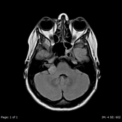
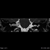
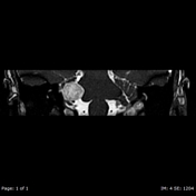
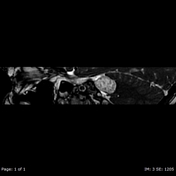
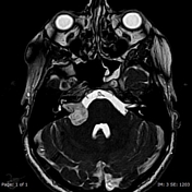
There is a complex mass arise from anatomic site of VIII parbased within the right cerebellar-pontine angle, with enlargementwidening of the right IAC andinternal auditory canal, involvement of both cochlear and vestibular branches of CN VIII.
Images Changes:
Image MRI (T1) ( update )

Description
was changed:
There is a heterogeneous hipointensehypointense mass arise fromwithin the right cerebelar pedunclecerebellar-pontine angle.
Image MRI (FLAIR) ( update )
Description
was changed:
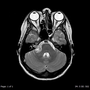
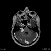
Image MRI (T2) ( update )
Description
was changed:
Heterogeneous hiperintensehyperintense on T2W with enlargement of right IAC
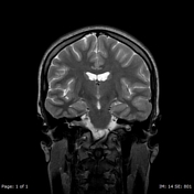
Image MRI (T2) ( update )
Description
was changed:
Image MRI (T2) ( update )

Description
was removed:
Image MRI (T2) ( update )
Description
was removed:
Image MRI (T2 fat sat) ( update )
Description
was changed:
IAC specific sequence nicely depictdepicts mass about CN VIII sheath
Image MRI (T2 fat sat) ( update )
Description
was changed:
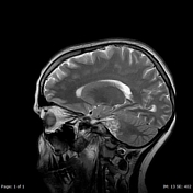
Well defined borders inadjacent to the right cerebelarcerebellar peduncle
Image MRI (T2 fat sat) ( update )
Description
was changed:
Well defined borders inadjacent to the right cerebelarcerebellar peduncle
Image MRI (T2 fat sat) ( update )
Description
was removed:
Image MRI (T1 C+) ( update )
Description
was changed:


 Unable to process the form. Check for errors and try again.