
Adrenal myelolipoma
Diagnosis almost certain
Updates to Case Attributes
Race
changed from Middle eastern to .
Diagnostic Certainty
was set to
.
Updates to Study Attributes
Findings
was added:
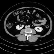
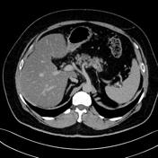
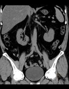
A small, heterogeneous, left adrenal, well defined lesion is seen. It has fat attenuation making up the majority, with a smaller, soft tissue attenuation in its center. No appreciable enhancement can be seen. Features highly representing left adrenal myelolipoma.
No retroperitoneal hemorrhage.
Diffuse fatty infiltration of the liver.
No further abnormality is seen in the abdominal solid viscera.
Images Changes:
Image CT (non-contrast) ( update )

Perspective
was set to
Axial.
Image CT (C+ arterial phase) ( update )

Perspective
was set to
Axial.
Image CT (C+ portal venous phase) ( update )
Perspective
was set to
Axial.
Image CT (C+ delayed) ( update )
Perspective
was set to
Axial.
Image CT (non-contrast) ( update )
Perspective
was set to
Coronal.
Updates to Study Attributes
Findings
was added:
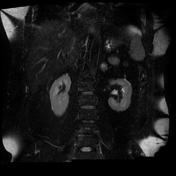
The out-of-phase images show striking drop of signal, mostly as a result of the lesion being of a mixed component (soft tissue and fat).
Images Changes:
Image MRI (T1 in-phase) ( update )

Perspective
was set to
Coronal.
Image MRI (T1 out-of-phase) ( update )

Perspective
was set to
Coronal.
Image MRI (T2 fat sat) ( update )
Perspective
was set to
Coronal.


 Unable to process the form. Check for errors and try again.