
Aortic valve stenosis
Diagnosis almost certain
Updates to Study Attributes
Images Changes:
Image X-ray (Frontal) ( update )
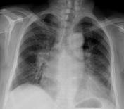
Perspective
was set to
Frontal.
Image X-ray (Lateral) ( update )
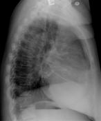
Perspective
was set to
Lateral.
Image 1 X-ray (Frontal) ( create )
Image 2 X-ray (Lateral) ( create )
Updates to Study Attributes
Images Changes:
Image CT (C+ portal venous phase) ( update )

Cropped
image
Perspective
was set to
Coronal.
Image CT (C+ portal venous phase) ( update )

Cropped
image
Perspective
was set to
Sagittal.
Image CT (C+ portal venous phase) ( update )
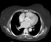
Perspective
was set to
Axial.
Cropped
image


 Unable to process the form. Check for errors and try again.