
Aortopulmonary window, interrupted aortic arch and large PDA giving the descending aorta
Updates to Case Attributes
Aortopulmonary window, (partial truncus arteriosus or aortopulmonary septal defect) is a rare congenital conotruncal cardiac anomaly with communication between the ascending aorta & the main or right pulmonary artery. It is caused by incomplete division of the embryonic common trunk. The two semilunar valves are usually separately identified and the absence of VSD differentiates it from truncus arteriosus. Association with PDA in 15%. Aortopulmonary window is associated with VSD or aortic coarctation.
In this case, interrupted aortic arch is seen with a large PDA is giving the descending aorta.
The Case is courtesy of Dr. Mohammad A. ElBeialy, MD, FRCR & Dr. Heba Kamal.
-<p><a href="/articles/aortopulmonary-window">Aortopulmonary window</a>, (partial <a href="/articles/truncus-arteriosus">truncus arteriosus</a> or <a href="/articles/aortopulmonary-septal-defect">aortopulmonary septal defect</a>) is a rare congenital <a href="/articles/conotruncal-cardiac-anomalies">conotruncal cardiac anomaly</a> with communication between the ascending aorta & the main or right pulmonary artery. It is caused by incomplete division of the embryonic common trunk. The two semilunar valves are usually separately identified and the absence of VSD differentiates it from truncus arteriosus. Association with <a href="/articles/patent-ductus-arteriosus">PDA</a> in 15%. Aortopulmonary window is associated with <a href="/articles/ventricular-septal-defect-1">VSD</a> or aortic coarctation.</p><p>In this case, <a href="/articles/interrupted-aortic-arch">interrupted aortic arch</a> is seen with a large PDA is giving the descending aorta.</p><p>The Case is courtesy of Dr. Mohammad A. ElBeialy, MD, FRCR & Dr. Heba Kamal.</p>- +<p><a href="/articles/aortopulmonary-window-radiograph">Aortopulmonary window</a>, (partial <a href="/articles/truncus-arteriosus">truncus arteriosus</a> or <a href="/articles/aortopulmonary-septal-defect">aortopulmonary septal defect</a>) is a rare congenital <a href="/articles/conotruncal-cardiac-anomalies">conotruncal cardiac anomaly</a> with communication between the ascending aorta & the main or right pulmonary artery. It is caused by incomplete division of the embryonic common trunk. The two semilunar valves are usually separately identified and the absence of VSD differentiates it from truncus arteriosus. Association with <a href="/articles/patent-ductus-arteriosus">PDA</a> in 15%. Aortopulmonary window is associated with <a href="/articles/ventricular-septal-defect-1">VSD</a> or aortic coarctation.</p><p>In this case, <a href="/articles/interrupted-aortic-arch">interrupted aortic arch</a> is seen with a large PDA is giving the descending aorta.</p><p> </p><p>The Case is courtesy of Dr. Mohammad A. ElBeialy, MD, FRCR & Dr. Heba Kamal.</p>
References changed:
- 1. Costa TJ, Damry N, Jacquemart C et-al. Aortopulmonary window: a rare congenital heart disease. JBR-BTR.97 (6): 356-7. <a href="http://www.ncbi.nlm.nih.gov/pubmed/25786293">Pubmed citation</a><span class="auto"></span>
- 2. Rider OJ, Bissell M, Myerson SG. Congenital aortopulmonary window; an unusual cause of breathlessness. Heart. 2013;99 (20): 1546. <a href="http://dx.doi.org/10.1136/heartjnl-2013-303753">doi:10.1136/heartjnl-2013-303753</a> - <a href="http://www.ncbi.nlm.nih.gov/pubmed/23735938">Pubmed citation</a><span class="auto"></span>
- 1- Costa TJ, Damry N, Jacquemart C et-al. Aortopulmonary window: a rare congenital heart disease. JBR-BTR.97 (6): 356-7. <a href="http://www.ncbi.nlm.nih.gov/pubmed/25786293">Pubmed citation</a><span class="auto"></span>
- 2- Rider OJ, Bissell M, Myerson SG. Congenital aortopulmonary window; an unusual cause of breathlessness. Heart. 2013;99 (20): 1546. <a href="http://dx.doi.org/10.1136/heartjnl-2013-303753">doi:10.1136/heartjnl-2013-303753</a> - <a href="http://www.ncbi.nlm.nih.gov/pubmed/23735938">Pubmed citation</a><span class="auto"></span>
Systems changed:
- Vascular
Updates to Study Attributes
-
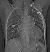
Athe scout revealed mild cardiomegaly with mild left ventricular dilatation and dilated aorta with increased pulmonary vascularity; a finding indistinguishable from isolated PDA. -
a relatively large aortopulmonary window is seen connecting the ascending aorta (about 1.5 cm distal to the aortic annulus) & the main pulmonary artery. The aorta is seen arising from the left ventricle and the ascending aorta ends by giving three orderly branches (the left subclavian, the left common carotid and brachiocephalic arteries)
. -
Interruptedinterrupted aortic arch is seen with a large PDA arising from the anterosuperior aspect of the distal main pulmonary artery and continues as the descending aorta in the left paravetebral gutter. No evident abdominal coarctation. -
Antegradeantegrade continuity between the right ventricle and the main pulmonary artery. Confluent dilated main pulmonary artery and its branches. -
Situssitus solitus, with the cardiac apex to the left. -
Mildmild cardiomegaly is noted with mild left ventricular dilatation. -
Atrioatrio-ventricular concordance and ventriculo-arterial concordance. Intactintact interatrial septum (IAS) & interventricular septum (IVS).-
Normalnormal origin and distribution of the coronaries. -
Increasedincreased pulmonary vascularity. No evidence of anomalous pulmonary venous drainage.
Image CT (Scout) ( update )
Updates to Study Attributes
Aortopulmonary window, interrupted aortic arch & large PDA giving the descending aorta..


 Unable to process the form. Check for errors and try again.