
Cerebral abscess - shaggy borders
Diagnosis almost certain
Updates to Study Attributes
Findings
was changed:
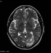
MRI of the brain demonstrates, within the left occipital lobe, a rounded region demonstrating central high T2, low T1 signal is demonstrated. It is bounded by a region of relative low signal on T2 and surrounded by extensive vasogenic oedema.
Diffusion weighted-weighted imaging demonstrates marked restriction, and the periphery of the lesion demonstrated bright ring ring enhancement with with somewhat shaggy borders.
MRS (performed in of the lesion(TE = 144 ) has a noisy base linebaseline but an inverted lactate lactate peak is is none the less visible, as are reduced choline, creatine, and NAA peaks.
Findings are consistent with a cerebral cerebral abscess.
Images Changes:
Image MRI (T2) ( update )
Perspective
was set to
Axial.
Single Or Stack Root
was set to
.
Image MRI (DWI) ( update )

Perspective
was set to
Axial.
Single Or Stack Root
was set to
.
Image MRI (T1) ( update )

Perspective
was set to
Axial.
Single Or Stack Root
was set to
.
Image MRI (T1 C+) ( update )

Perspective
was set to
Axial.
Single Or Stack Root
was set to
.
Image MRI (T1 C+) ( update )

Perspective
was set to
Coronal.
Single Or Stack Root
was set to
.
Image MRI (T1 C+) ( update )

Perspective
was set to
Sagittal.
Single Or Stack Root
was set to
.
Image MRI (MRS) ( update )

Description
was removed:
Single Or Stack Root
was set to
.
Updates to Case Attributes
Diagnostic Certainty
was set to
.


 Unable to process the form. Check for errors and try again.