
Cases
By sharing our collective experience through interesting and classic patient cases, we can make a real difference in how people are imaged and diagnosed. Each case belongs to a contributing member and all cases are reviewed by our dedicated editors to ensure they reach quality standards and abide by privacy guidelines. Cases can public or unlisted and then be viewed directly or added to articles, playlists or multiple choice questions. Find out more about cases.


205 results found
Case
Hypertrophic olivary degeneration (HOD)

Published
26 Jun 2023
92% complete
Diagram
MRI
Case
Pulmonary hamartoma

Published
29 May 2023
70% complete
Diagram
CT
Case
Anteroposterior compression II injury

Published
18 Apr 2023
69% complete
Diagram
X-ray
Case
Lateral compression 2 pelvic fracture

Published
18 Apr 2023
98% complete
X-ray
CT
Diagram
Case
Vertical shear injury

Published
17 Apr 2023
97% complete
X-ray
Diagram
Case
Skinfold mimics pneumothorax

Published
06 Apr 2023
91% complete
X-ray
Diagram
Case
Pancreatic pseudocyst with rupture

Published
16 Feb 2023
77% complete
Diagram
CT
Case
Morton neuroma (MRI)

Published
12 Feb 2023
77% complete
Diagram
MRI
Case
Ritalin lung

Published
03 Feb 2023
63% complete
X-ray
Diagram
Case
Anterior talofibular ligament injury with variant anatomy

Published
09 Dec 2022
69% complete
Ultrasound
Diagram
Annotated image
Case
Superior gluteal artery injury due to anteroposterior 2 compression injury

Published
07 Dec 2022
95% complete
DSA (angiography)
X-ray
CT
Diagram
Case
Appendicitis (CT angiogram)

Published
17 Oct 2022
65% complete
CT
Diagram
Case
Pacer pads and automated implantable cardioverter defibrillator in intubated patient

Published
22 Sep 2022
88% complete
Diagram
X-ray
Case
Median nerve injury

Published
16 Sep 2022
72% complete
Annotated image
Diagram
Ultrasound
Case
Fungal sinusitis
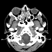
Published
11 Sep 2022
77% complete
CT
Diagram
Case
Four lumbar type vertebrae

Published
04 Sep 2022
84% complete
CT
Diagram
Case
Migration of supraspinatus tendon calcification

Published
15 Aug 2022
75% complete
Diagram
Ultrasound
Case
Osteomyelitis - femur
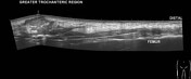
Published
24 Jul 2022
79% complete
Ultrasound
Diagram
X-ray
Case
Bullous emphysema

Published
30 Jun 2022
92% complete
X-ray
Diagram
CT
Case
Left main coronary artery arising from the right pulmonary artery

Published
15 Mar 2022
87% complete
X-ray
CT
MRI
Diagram
ADVERTISEMENT: Supporters see fewer/no ads


 Unable to process the form. Check for errors and try again.