
Cases
By sharing our collective experience through interesting and classic patient cases, we can make a real difference in how people are imaged and diagnosed. Each case belongs to a contributing member and all cases are reviewed by our dedicated editors to ensure they reach quality standards and abide by privacy guidelines. Cases can public or unlisted and then be viewed directly or added to articles, playlists or multiple choice questions. Find out more about cases.


181 results found
Case
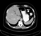
Mesenteric panniculitis and urolithiasis

Published
25 Jun 2010
83% complete
CT
Annotated image
Case
CT abdomen/pelvis coronal - labeling questions

Published
04 Dec 2018
40% complete
Annotated image
CT
Case
Wilms tumor: claw sign

Published
29 Jun 2013
98% complete
Annotated image
X-ray
CT
Case
Post-transplant lymphoproliferative disorder (small bowel)

Published
30 Aug 2009
95% complete
Annotated image
CT
Case
Transabdominal pelvic US - labeling questions

Published
06 Mar 2024
36% complete
Ultrasound
Annotated image
Case
Sex reassignment surgery
Published
30 Oct 2020
62% complete
Annotated image
CT
Case
Transitional cell carcinoma of the ureter

Published
17 Jan 2019
92% complete
Annotated image
CT
Ultrasound
Case
Pelviureteric junction obstruction with aberrant accessory lower pole renal artery

Published
31 Dec 2018
80% complete
Annotated image
CT
Case
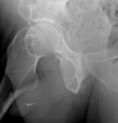
Migrated double J stent

Published
04 May 2018
91% complete
Annotated image
X-ray
Case
Urinary bladder hematoma

Published
05 May 2018
77% complete
Annotated image
Ultrasound
CT
Case
Urethral diverticulum (VCUG)

Published
12 Jan 2016
91% complete
Fluoroscopy
Annotated image
Case
Xanthogranulomatous pyelonephritis

Published
01 Jun 2010
96% complete
X-ray
Annotated image
MRI
CT
Case
Acute unilateral nonhemorrhagic adrenal infarction

Published
07 May 2019
77% complete
CT
Annotated image
Case
Vesico-vaginal fistula with ureteroureterostomy

Published
30 Mar 2017
95% complete
Fluoroscopy
CT
Annotated image
X-ray
Case
Wilms tumor

Published
20 Apr 2010
98% complete
Nuclear medicine
CT
Annotated image
Case
Renal infarction

Published
25 Mar 2010
52% complete
Annotated image
CT
Case
Tubular ectasia of the epididymus

Published
07 Aug 2022
79% complete
Ultrasound
Annotated image
Case
Adrenal glands (normal CT anatomy)

Published
29 Jan 2012
27% complete
Annotated image
CT
Case
Vesicoureteric junction stone

Published
20 Apr 2017
86% complete
Annotated image
X-ray
Case
Urethral glands of Littré
Published
26 Jun 2021
94% complete
Fluoroscopy
Annotated image
ADVERTISEMENT: Supporters see fewer/no ads


 Unable to process the form. Check for errors and try again.