
Items tagged “knee”
419 results found
Article
Anterior cruciate ligament mucoid degeneration
Anterior cruciate ligament (ACL) mucoid degeneration, along with tears and anterior cruciate ligament ganglion cysts, is a relatively common cause of increased signal within the anterior cruciate ligament (ACL). The appearance can mimic acute or chronic interstitial partial tears of the ACL. How...
Article
Anterior cruciate ligament ganglion cyst
Anterior cruciate ligament (ACL) ganglion cysts, commonly referred to simply as ACL cysts, along with ganglion cysts arising from the alar folds that cover the infrapatellar fat pad, make up the vast majority of intra-articular ganglion cysts of the knee.
Epidemiology
Anterior cruciate ligamen...
Article
Celery stalk sign (anterior cruciate ligament)
The celery stalk sign is a term given to the appearance of the anterior cruciate ligament which has undergone mucoid degeneration and has been likened to that of a celery stalk. Its low signal longitudinal fibers are separated from each other by higher signal mucinous material, best appreciated ...
Case
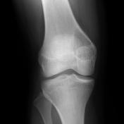
Sinding-Larsen-Johansson disease

Published
06 Jun 2009
58% complete
MRI
X-ray
Case
Lipohemarthrosis

Published
23 Aug 2009
69% complete
X-ray
Annotated image
Article
Pellegrini-Stieda lesion
Pellegrini-Stieda lesions refer to ossified post-traumatic lesions at (or near) the medial femoral collateral ligament adjacent to the margin of the medial femoral condyle. One presumed mechanism of injury is a Stieda fracture (avulsion injury of the medial collateral ligament at the medial femo...
Case
Insall-Salvati ratio - normal patella

Published
10 Sep 2009
39% complete
MRI
Case
Normal Insall-Salvati ratio (annotated image)

Published
10 Sep 2009
24% complete
MRI
Case
Chronic patellar tendinosis - Jumper's knee

Published
14 Sep 2009
59% complete
MRI
Case
Spontaneous osteonecrosis of the knee

Published
14 Sep 2009
64% complete
X-ray
MRI
Article
Absent bow tie sign (knee)
The absent bow tie sign represents the loss of the normal appearance of the menisci on parasagittal MRI images and is suggestive of meniscal injury.
Normally the medial and lateral menisci appear as low signal triangular structures linked by a thin body located between the femoral condyles and ...
Article
Fragment-in-notch sign (knee)
The fragment-in-notch sign is closely related to, can be thought of as a failed, double PCL sign, in so far as it represents a meniscal fragment lying in the intercondylar notch, but not paralleling the posterior cruciate ligament (PCL).
Although both signs are most frequently seen in the sett...
Article
Posterior cruciate ligament
The posterior cruciate ligament (PCL) is one of the two cruciate ligaments that stabilize the knee joint.
Gross anatomy
The posterior cruciate ligament arises from the lateral surface of the medial femoral condyle and inserts onto the posterior lateral intercondylar surface of the tibial plat...
Case
Bucket handle tear - lateral meniscus

Published
22 Sep 2009
74% complete
MRI
Case
Lateral patellar dislocation

Published
23 Sep 2009
74% complete
MRI
Case
Patellar dislocation

Published
22 Oct 2009
53% complete
MRI
Case
Ossified subperiosteal hematoma - femur

Published
03 Nov 2009
41% complete
X-ray
Case
Giant cell tumor - atypical
Published
04 Nov 2009
84% complete
CT
X-ray
MRI
Article
Patella baja
Patella baja, also known as patella infera, is an abnormally low lying patella, which is associated with restricted range of motion, crepitations, and retropatellar pain. If longstanding, extensor dysfunction may ensue with significant morbidity.
Pathology
It is seen in a variety of clinical s...
Article
Blackburne-Peel ratio
The Blackburne-Peel ratio is an alternative to Insall-Salvati ratio and is used to assess patellar height including patella alta and baja.
Usage
The Blackburne-Peel ratio has higher interobserver reliability compared to the Insall-Salvati ratio and can also be used when the tibial tuberosity ...


 Unable to process the form. Check for errors and try again.