


44 results found
Case
Apophyseal avulsion fractures of the pelvis and hip (illustration)

Published
01 Apr 2024
44% complete
Diagram
Case
Blood supply of the femoral head (illustration)

Published
05 Feb 2024
44% complete
Diagram
Case
Slipped capital femoral epiphysis (illustrations)

Published
26 Dec 2023
41% complete
Diagram
Case
Rectal anatomy (illustrations)

Published
24 May 2023
41% complete
Diagram
Case
Pediatric hand anatomy (illustration)

Published
24 May 2023
35% complete
Diagram
Case
Lauge-Hansen classification of ankle fractures (illustrations)

Published
01 Nov 2022
36% complete
Diagram
Case
Ankle tear drop sign (illustration)

Published
01 Nov 2022
41% complete
Diagram
Case
Chopart and Lisfranc joints (illustrations)

Published
29 Aug 2022
41% complete
Diagram
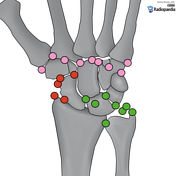
Case
Carpal dislocations (illustrations)

Published
09 Aug 2022
38% complete
Diagram
Case
Nunley-Vertullo classification of Lisfranc injuries (illustrations)

Published
04 May 2022
41% complete
Diagram
Case
Lisfranc injury - Myerson classifications (illustrations)

Published
04 May 2022
35% complete
Diagram
Case
Lisfranc ligamentous complex (illustration)

Published
03 May 2022
44% complete
Diagram
Case
Regions of the foot (illustrations)

Published
02 May 2022
44% complete
Diagram
Case
Accessory ossicles of the foot (illustration)

Published
02 May 2022
44% complete
Diagram
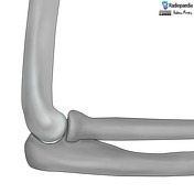
Case
Elbow anatomy (illustration)

Published
18 Apr 2022
35% complete
Diagram
Case
Supracondylar fracture (illustrations)

Published
14 Feb 2022
35% complete
Diagram
Case
Pediatric elbow anatomy (illustrations)

Published
08 Feb 2022
35% complete
Diagram
Case
Milch classification of lateral humeral condyle fractures (illustrations)

Published
08 Feb 2022
44% complete
Diagram
Case
Patterns of knee injury

Published
18 Oct 2021
41% complete
Diagram
Case
Knee joint dislocations (illustrations)

Published
17 Oct 2021
36% complete
Diagram
Case
AO classification of distal femur fractures (illustration)
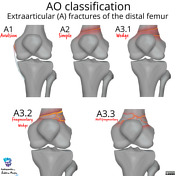
Published
17 Oct 2021
44% complete
Diagram
Case
Schatzker classification of tibial plateau fractures (illustrations)

Published
09 Oct 2021
35% complete
Diagram
Case
Knee radiograph checklist (illustration)

Published
08 Oct 2021
44% complete
Diagram
Case
Lipohemarthrosis and hemarthrosis (illustrations)

Published
08 Oct 2021
44% complete
Diagram
Case
Knee anatomy (illustrations)

Published
04 Oct 2021
35% complete
Diagram
Case
Mallet finger (illustration)

Published
22 Apr 2021
41% complete
Diagram
Case
Elbow joint effusion (illustration)
Published
08 Feb 2021
35% complete
Diagram
Case
AO/OTA classification of distal humeral fractures

Published
15 Jan 2021
35% complete
Diagram
Case
Scapholunate advanced collapse (illustration)

Published
14 Aug 2020
32% complete
Diagram
Case
Scaphoid non-union advanced collapse (illustration)

Published
12 Aug 2020
32% complete
Diagram
Case
Radial height (illustration)

Published
11 Aug 2020
41% complete
Diagram
Case
Normal wrist alignment, dorsal and volar intercalated segmental instability (illustration)

Published
10 Aug 2020
35% complete
Diagram
Case
Ossicles of the wrist and hand (illustration)
Published
10 Aug 2020
29% complete
Diagram
Case
Wrist anatomy (illustration)

Published
10 Aug 2020
29% complete
Diagram
Case
Lateral wrist anatomy (illustration)

Published
09 Aug 2020
35% complete
Diagram
Case
Rockwood classification of acromioclavicular joint injury

Published
25 Nov 2019
22% complete
Diagram
Case
Images for MCQs

Published
08 Oct 2019
18% complete
Diagram
Case
Recurrent neural network

Published
05 Jul 2019
22% complete
Diagram
Case
Simplified neural network

Published
09 Jun 2019
35% complete
Diagram
Case
Convolutional neural network (diagram)

Published
01 May 2019
44% complete
Diagram
Case
TS-OP line (diagram)

Published
02 Oct 2018
25% complete
Diagram
Case
Skull positioning lines (diagram)

Published
03 Mar 2018
38% complete
Diagram
Case
Paravertebral gutter technique (diagram)

Published
31 Jul 2017
41% complete
Diagram
Case
Iodinated contrast (diagram)
Published
15 Oct 2016
32% complete
Diagram
ADVERTISEMENT: Supporters see fewer/no ads


 Unable to process the form. Check for errors and try again.