
Cases
By sharing our collective experience through interesting and classic patient cases, we can make a real difference in how people are imaged and diagnosed. Each case belongs to a contributing member and all cases are reviewed by our dedicated editors to ensure they reach quality standards and abide by privacy guidelines. Cases can public or unlisted and then be viewed directly or added to articles, playlists or multiple choice questions. Find out more about cases.


1,701 results found
Case
Anterior talofibular ligament injury with variant anatomy

Published
09 Dec 2022
69% complete
Ultrasound
Diagram
Annotated image
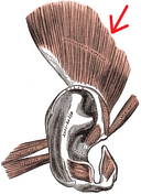
Case
Anterior auricular muscle (Gray's illustration)

Published
27 Jun 2023
29% complete
Diagram
Case
Pathological fracture of enchondroma

Published
15 Jan 2020
79% complete
X-ray
Diagram
Case
Anterior auricular muscle (Gray's illustration)

Published
27 Jun 2023
29% complete
Diagram
Case
Splenic injury - AAST grade III

Published
10 Nov 2015
100% complete
CT
Diagram
Case
Pes anserinus tendon (diagram)

Published
26 Feb 2012
25% complete
Annotated image
Diagram
Case
Abducens and facial cranial nerves and nuclei
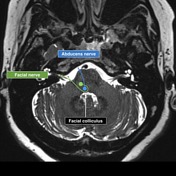
Published
23 Jan 2017
32% complete
Diagram
Case
The yin-yang sign

Published
11 Mar 2012
25% complete
Diagram
Case
Superior auricular muscle (Gray's illustration)
Published
27 Jun 2023
29% complete
Diagram
Case
Echinococcus life cycle (diagram)
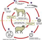
Published
15 May 2015
29% complete
Diagram
Case
Development of double aortic arch (illustration)

Published
10 Oct 2018
32% complete
Diagram
Case
Chiari 1.5 malformation

Published
13 Jan 2017
87% complete
Diagram
MRI
Case
Superior auricular muscle (Gray's illustration)

Published
27 Jun 2023
29% complete
Diagram
Case
Ascending spinal tracts (Gray's illustration)

Published
08 Sep 2020
35% complete
Diagram
Case
Bone-RADS flowcharts
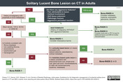
Published
28 Apr 2022
29% complete
Diagram
Case
Cranial nerves in the posterior fossa (Gray's illustration)

Published
06 Sep 2020
35% complete
Diagram
Case
Wernicke encephalopathy

Published
16 Feb 2022
80% complete
MRI
Diagram
Case
Chondrocalcinosis: diagram of causes

Published
01 Oct 2012
32% complete
Diagram
Case
True bovine arch (illustration)

Published
10 May 2015
22% complete
Diagram
Case
Platybasia (diagrams)

Published
07 May 2008
32% complete
Diagram
ADVERTISEMENT: Supporters see fewer/no ads


 Unable to process the form. Check for errors and try again.