


7,872 results found
Case
Normal AP mandible radiograph

Published
06 Jul 2016
19% complete
X-ray
Case
Bilateral cleft palate

Published
05 Jul 2016
75% complete
X-ray
Article
Intrinsic muscles of the larynx
The intrinsic muscles of the larynx can be considered in two groups:
muscles that control the inlet of the larynx
muscles that move the vocal ligaments
Gross anatomy
Muscles of the inlet
aryepiglottic muscle: lies within the aryepiglottic fold, runs from the side of the epiglottis and inser...
Case
Phthisis bulbi

Published
02 May 2015
65% complete
CT
Case
Hat in the neck (Rorschach radiology)

Published
06 Nov 2016
36% complete
CT
Article
Sialolithiasis
Sialolithiasis refers to the formation of calculi (sialoliths) inside the ducts or parenchyma of salivary glands and most commonly occurs in the submandibular glands and their ducts.
Epidemiology
Sialolithiasis is the most common disease of salivary glands, accounting for approximately 50% of ...
Article
Aicardi syndrome
Aicardi syndrome is a rare severe developmental disorder. It results from an X-linked genetic defect that is fatal in males and therefore only manifests in females (except for rare 47, XXY cases).
Terminology
Aicardi syndrome is distinct from Aicardi-Goutieres syndrome although both are named ...
Article
Nasal dermoid cyst
Nasal dermoids (or nasal dermoid sinus cysts) are the most common congenital midline nasal lesion typically presenting in early childhood.
Epidemiology
Nasal dermoids are rare and account for only 4-12% of all dermoid cysts of the head and neck, far less common than angular dermoids 1,2. They ...
Article
Frontal intersinus septal cells
Frontal intersinus septal cells, also known as interfrontal sinus septal cells, are a subtype of medial frontal recess cells.
Gross anatomy
The frontal intersinus septal cells lie within the intersinus septum between the frontal sinuses. They usually drain in the medial aspect of the frontal r...
Article
Retromolar trigone
The retromolar trigone, sometimes called the retromolar fossa, is an oral cavity subsite that consists of the mucosa posterior to the last mandibular molar. It is roughly triangular shaped and extends superiorly towards the maxilla along the anterior surface of the mandible.
Gross anatomy
Att...
Article
Kayser-Fleischer rings
Kayser-Fleischer rings, sometimes shortened to K-F rings, are caused by copper deposition in the cornea and are a specific, clinical sign of Wilson disease.
Clinical presentation
They are usually brown or dark reddish in color. Early on they may need a slit lamp to be visible before they becom...
Article
Deep spaces of the head and neck
The deep spaces of the head and neck refer to compartments delimited by the deep cervical fascia. While these concepts overlap with traditional anatomical descriptions, their existence highlights the importance of fascia in confining various pathologies.
A knowledge of these spaces not only all...
Case
Metastatic melanoma to the orbit

Published
16 May 2023
95% complete
CT
MRI
Case
Venous malformation of the face
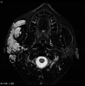
Published
25 Jul 2008
71% complete
MRI
CT
Article
Primary acquired nasolacrimal duct obstruction (PANDO)
Primary acquired nasolacrimal duct obstruction (PANDO) is a chronic inflammatory cause of nasolacrimal drainage apparatus obstruction.
Epidemiology
Most commonly seen in middle-aged or elderly women.
Pathology
Etiology
The exact cause is still not very well known however it thought to be...
Case
Osteolytic skull metastasis of lung cancer

Published
05 May 2013
89% complete
CT
Article
Anderson and Montesano classification of occipital condyle fractures
The Anderson and Montesano classification is a widely used system for describing occipital condyle fractures. It divides injuries into three types based on morphology and mechanism of injury 1-5.
Classification
type I: impacted type occipital condyle fracture
morphology: comminution of the co...
Case
Juvenile nasopharyngeal angiofibroma

Published
10 Nov 2014
89% complete
MRI
Article
Orthopantomography
The orthopantomogram (also known as an orthopantomograph, pantomogram, OPG or OPT) is a panoramic single image radiograph of the mandible, maxilla and teeth. It is often encountered in dental practice and occasionally in the emergency department; providing a convenient, inexpensive and rapid way...
Case
Chordoma - clivus
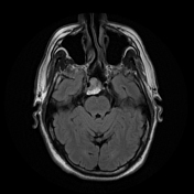
Published
18 Jan 2022
88% complete
CT
MRI


 Unable to process the form. Check for errors and try again.