


1,812 results found
Article
Fetal right ventricular enlargement
Fetal right ventricular (RV) enlargement is an infrequently encountered situation in antenatal imaging.
Pathology
The right ventricle is the dominant ventricle during in utero development. Right ventricular enlargement can occur with a number of cardiac as well as non-cardiac anomalies.
cardi...
Case
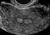
Uterine adhesion band
Published
04 Aug 2011
85% complete
Ultrasound
Case
Bilateral adrenal vein thrombosis

Published
05 Feb 2018
73% complete
CT
MRI
Ultrasound
Article
Microgenia
Microgenia is a term meaning a small chin. It is somewhat related to but distinct from the term micrognathia which means a small mandible.
Pathology
Associations
Microgenia can be isolated or be associated with a number of anomalies which include
campomelic dysplasia 1
hydrolethalus
Noonan...
Case
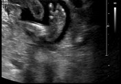
Live extrauterine ectopic pregnancy

Published
26 Aug 2020
77% complete
Ultrasound
MRI
Article
Early structural scan
An early structural scan is a first trimester obstetric ultrasound performed around 12-16 weeks gestation. It can have some similarity to the 11-13 week antenatal ultrasound and assess limited fetal anatomy. However, a formal evaluation of nuchal translucency is not part of this ultrasound asses...
Article
Fetal intra-abdominal cysts (differential)
Fetal intra-abdominal cystic lesions can arise from a number of physiological and pathological causes.
Physiological
fetal gastric dilatation / fetal gastric bubble (can be pathological if there is a gastric outlet obstruction
normal fetal gallbladder
Pathological
No color flow
fetal chole...
Case
Triple loop nuchal cord

Published
06 Apr 2022
94% complete
Ultrasound
Case
Umbilical cord cysts

Published
03 Jul 2019
75% complete
Ultrasound
Case
Subchorionic hemorrhage

Published
28 Nov 2020
94% complete
Ultrasound
Case
Fetal rhombencephalosynapsis

Published
12 Apr 2017
77% complete
MRI
Case
Congenital talipes equinovarus - antenatal
Published
29 Nov 2020
72% complete
Ultrasound
Case
Failed early pregnancy

Published
04 Jul 2022
94% complete
Ultrasound
Case
Torsion of dermoid cyst in a primigravid

Published
21 Nov 2021
80% complete
MRI
Case
Scalp hematoma types (diagram)

Published
30 Apr 2018
38% complete
Diagram
Case
Marginal cord insertion

Published
29 Sep 2023
97% complete
Ultrasound
Case
Variation in placental morphology: diagrams

Published
14 Apr 2011
32% complete
Diagram
Article
Fetal atrioventricular block
Fetal atrioventricular block is a form a fetal bradyarrhythmia often classified into
fetal partial atrioventricular block (PAVB)
fetal complete atrioventricular block (CAVB)
Epidemiology
Fetal atrioventricular block is considered rare finding with reported occurrences of around 1:11,000-20,0...
Article
Cystic renal dysplasia
Cystic renal dysplasia refers to a subgroup of congenital anomalies of the kidney and urinary tract characterized by the dysplastic renal parenchyma and formation of cysts. The most severe form is multicystic dysplastic kidney, in which functional renal parenchyma is absent and only undifferenti...
Article
The crisscross sign
The crisscross sign is a fetal ultrasound sign that describes the normal relationship between the ventricular outflows tracts of the fetal heart. The left ventricular outflow tract (LVOT); represented by the take-off of the aorta (Ao) from the left ventricle, is perpendicular (90o) to the right ...


 Unable to process the form. Check for errors and try again.