


463 results found
Case
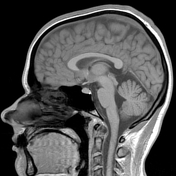
Creutzfeldt-Jakob disease
Published
28 May 2023
71% complete
MRI
Case
Posterior oblique ligament injury

Published
23 May 2021
71% complete
Annotated image
MRI
Case
Round atelectasis

Published
14 Mar 2014
71% complete
X-ray
CT
Case
Excessive lateral pressure syndrome

Published
18 Jul 2020
71% complete
Annotated image
MRI
Case
Foreign body granuloma (cactus spine)
Published
21 Mar 2022
71% complete
X-ray
Ultrasound
Case
Acromioclavicular joint injury - type V

Published
16 Mar 2021
71% complete
X-ray
Case
Pituicytoma

Published
03 Jul 2021
70% complete
MRI
Case
Pulmonary hamartoma

Published
23 Oct 2020
70% complete
Annotated image
CT
Case
Intradiploic nontraumatic sphenoid arachnoid pit

Published
15 Sep 2020
70% complete
CT
Annotated image
Case
Right middle lobe pneumonia

Published
03 Jun 2019
69% complete
X-ray
Case
Acromioclavicular joint injury - Rockwood I

Published
18 Feb 2019
69% complete
Annotated image
X-ray
Case
Respiratory distress syndrome (severe)

Published
13 Jul 2018
69% complete
X-ray
Case
Interphalangeal joint dislocation of the hallux

Published
07 Nov 2020
69% complete
X-ray
Case
Avascular necrosis of the femoral head

Published
18 Jul 2020
69% complete
X-ray
Case
Os subfibulare

Published
24 Feb 2014
69% complete
X-ray
Case
Ligamentum mucosum injury

Published
01 May 2016
68% complete
MRI
Case
Thalamic glioma

Published
23 May 2021
68% complete
MRI
Case
Subacute pontine infarct

Published
11 Jun 2022
68% complete
CT
Case
Twinkling artifact caused by calcified renal cyst

Published
22 Nov 2018
68% complete
Ultrasound
Case
Splenic hamartoma

Published
21 Aug 2020
68% complete
CT
Case
Tolosa-Hunt syndrome
Published
14 Nov 2021
68% complete
MRI
Case
Renal angiomyolipoma

Published
03 Oct 2017
68% complete
Ultrasound
Case
Cerebral cavernous venous malformation (Zabramski type II)

Published
02 Jan 2021
67% complete
Annotated image
MRI
Case
Multiple blood blister-like supraclinoid internal carotid artery aneurysms
Published
27 Mar 2021
67% complete
MRI
Case
Chronic Kerley B lines in congestive heart failure

Published
04 Mar 2020
66% complete
X-ray
Case
Focal hepatic steatosis
Published
28 Nov 2018
66% complete
Ultrasound
Case
Wet lung and pulmonary venous congestion in the context of patent ductus arteriosus and patent foramen ovale

Published
21 Feb 2021
66% complete
X-ray
Case
Muscle hernia - rectus abdominis

Published
23 Feb 2017
66% complete
Ultrasound
Case
Unilateral hypoplastic first rib

Published
01 Jul 2018
66% complete
X-ray
Case
Buford complex

Published
01 Apr 2020
65% complete
Annotated image
MRI
Case
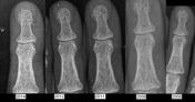
Pediatric olecranon fracture - subtle

Published
14 Dec 2018
65% complete
Annotated image
X-ray
Case
Intratendinous ganglion cyst of the Achilles tendon

Published
23 Aug 2021
64% complete
MRI
Case
Normal trauma protocol brain and C-spine CT

Published
15 Dec 2020
64% complete
CT
MRI
Case
Lipiodol lymphography (historical)

Published
03 Feb 2021
63% complete
X-ray
Case
Bilateral sacroiliitis - New York grade III
Published
14 Feb 2021
63% complete
X-ray
Case
Right middle lobe pneumonia (subtle)

Published
09 Jul 2020
63% complete
X-ray
Case
Ischiofemoral impingement

Published
15 Feb 2022
63% complete
MRI
Ultrasound
Annotated image
X-ray
Case
Subconjunctival fat prolapse
Published
20 Apr 2022
62% complete
MRI
Case
Ring artifact on low dose CT

Published
06 Jun 2021
62% complete
CT
Case
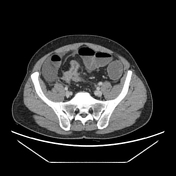
Perforated sigmoid diverticulitis

Published
03 Feb 2022
62% complete
CT
Case
Effect of iterative metal artifact reduction

Published
10 Jul 2022
62% complete
CT
Case
Sigmoid diverticulitis

Published
07 Feb 2019
61% complete
Annotated image
Ultrasound
Case
Multinodular and vacuolating neuronal tumor

Published
02 Nov 2019
61% complete
MRI
Annotated image
Case
Overexposed radiograph (chest x-ray)

Published
20 Jun 2019
60% complete
X-ray
Case
Hashimoto thyroiditis
Published
26 Oct 2020
60% complete
Ultrasound
Case
Radial styloid process fracture (pediatric)

Published
27 Oct 2019
60% complete
X-ray
Case
Normal dual-energy chest CT angiography with pulmonary embolism protocol

Published
14 Feb 2021
59% complete
CT
Case
Tonsilloliths

Published
13 Apr 2013
59% complete
CT
Case
Superior accessory fissure

Published
23 Aug 2019
59% complete
CT
Case
Striated nephrogram

Published
14 Mar 2016
59% complete
CT
Case
Double abdominal aorta artifact on ultrasound

Published
07 Dec 2020
59% complete
Ultrasound
CT
Annotated image
Case
Squamous cell cancer of the nasofacial angle

Published
04 Jun 2021
59% complete
CT
Case
Os cubiti anterior

Published
19 Jun 2020
58% complete
X-ray
Annotated image
Case
Lipid-rich adrenal adenoma

Published
29 Dec 2020
58% complete
Ultrasound
CT
Case
Focal hepatic steatosis - absent vessel distortion

Published
22 Jan 2020
57% complete
Ultrasound
Case
Intravenous cholecystography (historical)

Published
11 Dec 2019
57% complete
X-ray
Case
Normal noncontrast MR venography

Published
05 Apr 2021
56% complete
MRI
Case
Relationship between color Doppler flash artifact and color-write priority

Published
18 Feb 2020
54% complete
Ultrasound
Case
Otomastoiditis

Published
15 Dec 2015
53% complete
CT
Case
Normal duplex ultrasound of a transplant kidney

Published
27 Mar 2021
50% complete
Ultrasound
Case
Normal Waters view skull x-ray

Published
13 Sep 2018
50% complete
X-ray
Annotated image
Case
Physiologic age-related pituitary volume loss
Published
09 May 2021
50% complete
Annotated image
MRI
Case
Interzygomatic line

Published
20 Nov 2019
50% complete
CT
Case
Dual-chamber cardiac pacemaker

Published
04 Dec 2020
50% complete
X-ray
Case
Movement artifact (standing erect x-ray)

Published
26 Jun 2019
50% complete
Case
X-ray bronchography (historical)

Published
11 Dec 2019
50% complete
X-ray
Case
Bowel obstruction due to metastatic sigmoid tumor
Published
27 Jun 2022
48% complete
X-ray
CT
Case
Antibiotic spacer (hip)

Published
04 Jan 2021
47% complete
X-ray
Case
Spurious spectral broadening (ultrasound)

Published
05 Feb 2020
47% complete
Case
Platybasia

Published
03 Mar 2014
45% complete
CT
Case
Blooming artifact (CT)

Published
26 Aug 2019
45% complete
CT
Case
Normal B-flow portal venous flow

Published
19 Feb 2021
44% complete
Ultrasound
Case
Signal dropout on US probe due to dead element
Published
20 Dec 2020
44% complete
Ultrasound
Case
Chest x-ray (expiration and inspiration views)

Published
13 Sep 2017
44% complete
X-ray
Case
Rheumatoid arthritis - progressive changes in distal interphalangeal joint
Published
25 Jul 2014
44% complete
X-ray
Case
Normal paper clip test of an ultrasound probe

Published
02 Jan 2021
41% complete
Ultrasound
Case
Blooming artifact (ultrasound)

Published
01 Dec 2018
41% complete
Ultrasound
Case
Mandibula radiographs (lateral views)

Published
06 Oct 2020
41% complete
X-ray
Case
Delamination artifact on US probe

Published
20 Dec 2020
41% complete
Ultrasound
Case
CEUS appearance of Bosniak renal cystic masses (illustrations)
Published
31 Oct 2021
41% complete
Annotated image
Case
Negative trauma CT brain (pediatric)

Published
11 Jun 2022
39% complete
CT
Case
Flash mode (CEUS)

Published
17 Nov 2019
38% complete
Ultrasound
Case
Normal low-dose chest CT

Published
22 Sep 2021
36% complete
CT
Case
Normal in-air reverberation pattern - linear and curvilinear array transducers

Published
20 Dec 2020
35% complete
Ultrasound
Case
Bowel chainsaw (Rorschach radiology)

Published
14 May 2019
33% complete
MRI
Case
Normal scaphoid series
Published
07 Nov 2020
32% complete
X-ray
Case
Aliasing artifact (ultrasound)

Published
08 Dec 2018
32% complete
Ultrasound
Case
Normal in-air reverberation pattern - phased array transducer
Published
31 Dec 2020
29% complete
Ultrasound
Case
Multiple delamination defects on US probe

Published
02 Jan 2021
29% complete
Ultrasound
Case
Oligoprogression and polyprogression (illustration)

Published
29 Oct 2020
24% complete
Diagram
Case
Oligometastatic and polymetastatic disease (illustration)

Published
01 Nov 2020
24% complete
Diagram
ADVERTISEMENT: Supporters see fewer/no ads


 Unable to process the form. Check for errors and try again.